
Our case
History
1
Treatment Strategy
• Due to the small size of the aneurysm and the well-developed right PCA, we planned to embolize the aneurysm with simple coiling technique.
• If any loop protrusion during coiling ,stent-assisted coiling or Pcom artery sacrifice could be considered.
• Two Prowler Plus microcatheters were selected. One was for better supporting during coiling, the other was for ICA protection and stenting.
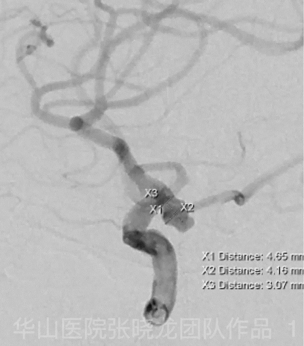
Figure 1. Measurement.
Video 2. The first Prowler Plus microcatheter was sent distal to the right MCA. The second Prowler Plus microcatheter with spiral curved tip was navigated to the aneurysm sac.
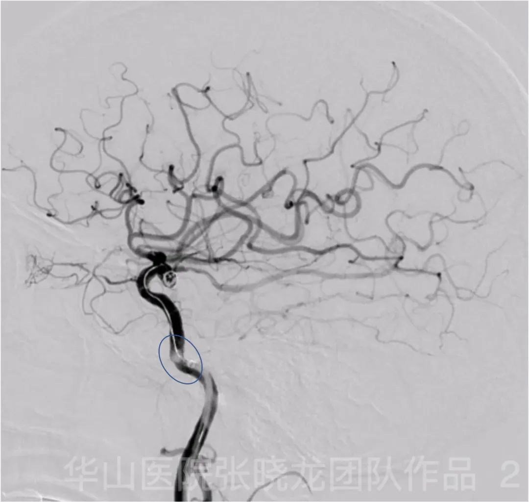
Figure 2. Angiography after inserting Perdenser 5mm*15cm coil for framing shows thrombosis in the right ICA. General Heparinization was administrated.

Figure 3. Roadmap shows the occlusion of right MCA, which is probable due to thrombus migration.

Figure 4. Perdenser 3mm*6mm and three Perdenser 3mm*4cm were inserted.

Figure 5 GIF. Perdenser 2mm*4cm was failed to inserted into the aneurysm sac.

Figure 6 GIF. Microcatheter passed the thrombus and was placed in distal branch and superselective angiography shows thrombous in the right MCA bifurcation migrated from ICA.

Figure 7 GIF. Solitaire 4mm*20mm was deployed for clot retrieval.

Figure 8 GIF. Angiography shows paritial recanalization of the superior trunk of the right MCA.

Figure 9 GIF. Tirofiban 6ml was administrated. Waiting for 8 mins.

Figure 10 GIF. Second clot retrieval.

Figure 11 GIF. Waiting for 8 mins.
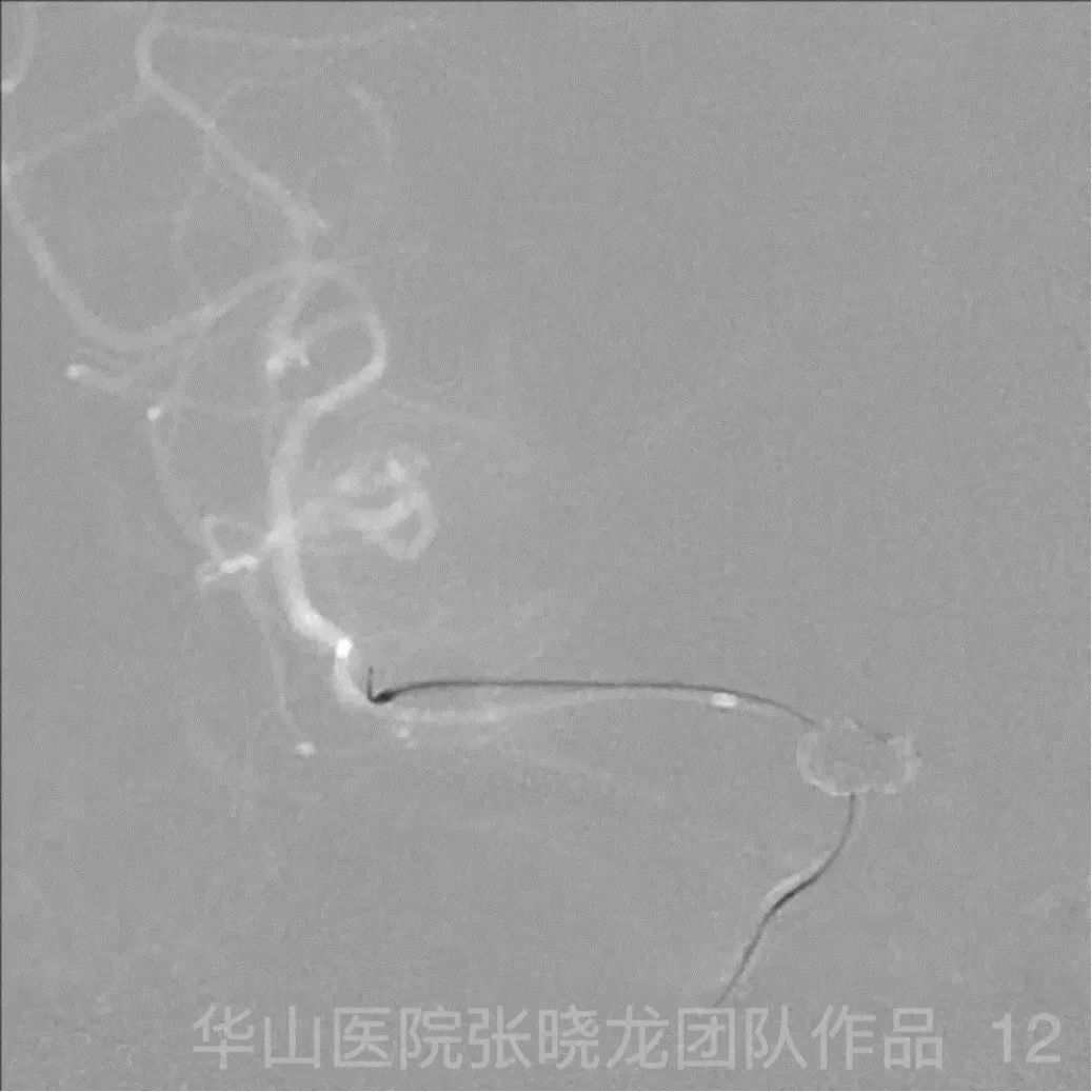
Figure 12 GIF. Third clot retrieval.

Figure 13 GIF. Tirofiban 3ml.

Figure 14 GIF. Angiography shows the densely packing of the aneurysm. The superior trunk of the right MCA is patent. Thrombosis is still noted in the initial segment of the inferior trunk of the right MCA,the blood flow is normal.

Figure 15 GIF. Post circulation angiography shows the patent of the right PCA and the pial compensation to the anterior circulation.

Figure 16 GIF. 72 hours CT shows right temporal lobe infarction.

Figure 17 GIF. The muscle strength of the left upper limb was V.

Figure 18 GIF. The muscle strength of the left lower limb was V.
2
Discharge
4
Summary
Thrombosis in the right ICA after inserting the first coil
Reason: The elder patient, two Prowler Plus microcatheters in a 6F guiding catheter without general heparinization may be the cause of right ICA thrombosis.
Treatment: General heparinization was given immediately. After observing the right MCA occlusion due to thrombus migration,clot retrieval was performed after densely coiling the aneurysm quickly.
Suggestions:
1. When the guiding was placed far enough (beyond the petrous segment),the risk of bleeding was reduced,heparinization should be performed.
2. If considering the risk of thrombosis,the combination of two microcatheters should be selected cautiously.
3. Avoid selecting two large size microcatheters.