Our case
History
• Female, 71-year-old.
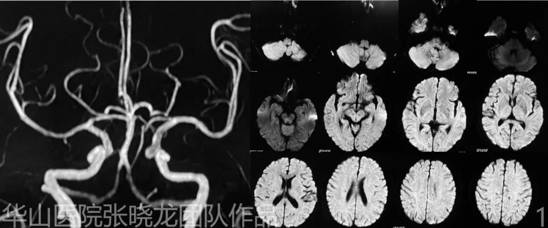
Figure 1. MRA shows a medial side of carotid-ophthalmic aneurysm. DWI shows no acute infarction.
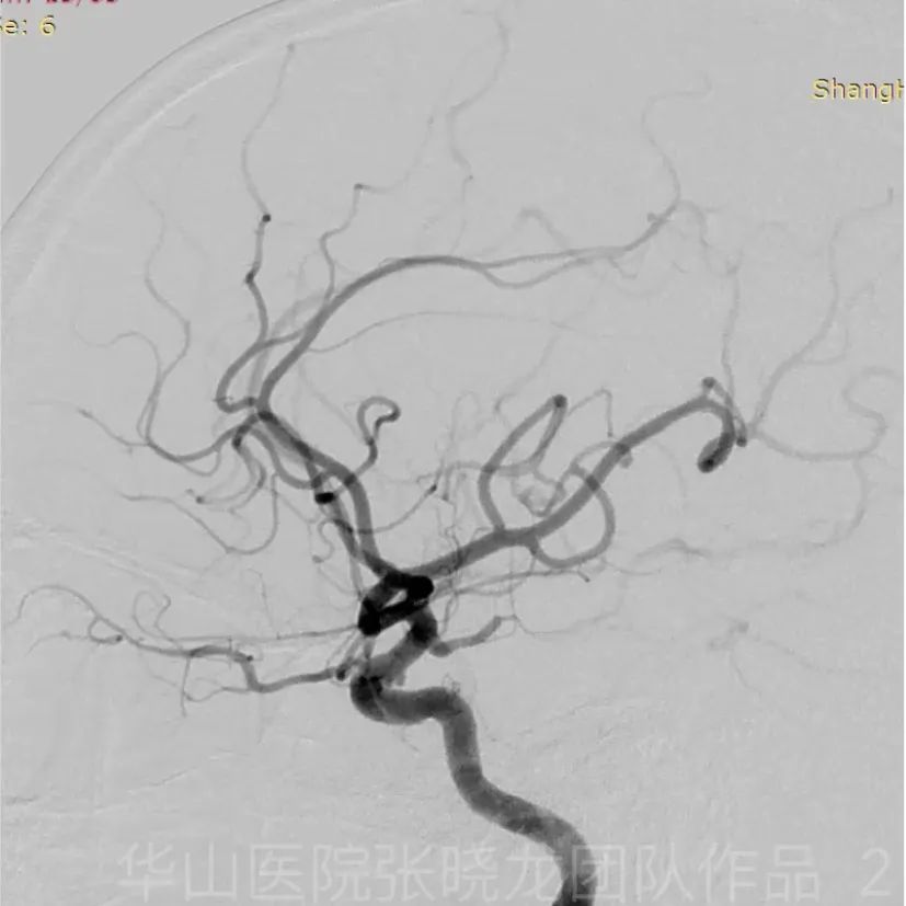
Figure 2. Two tiny aneurysms in ophthalmic segment of right ICA.
1
Strategy
• Two tandem ophthalmic aneurysms in the left ICA were revealed, one regular-shaped and wide-necked indicating a low rupture risk1, while the other is narrow-necked resulting in a high rupture risk1 which could be treated with simple coiling.
2
Operation
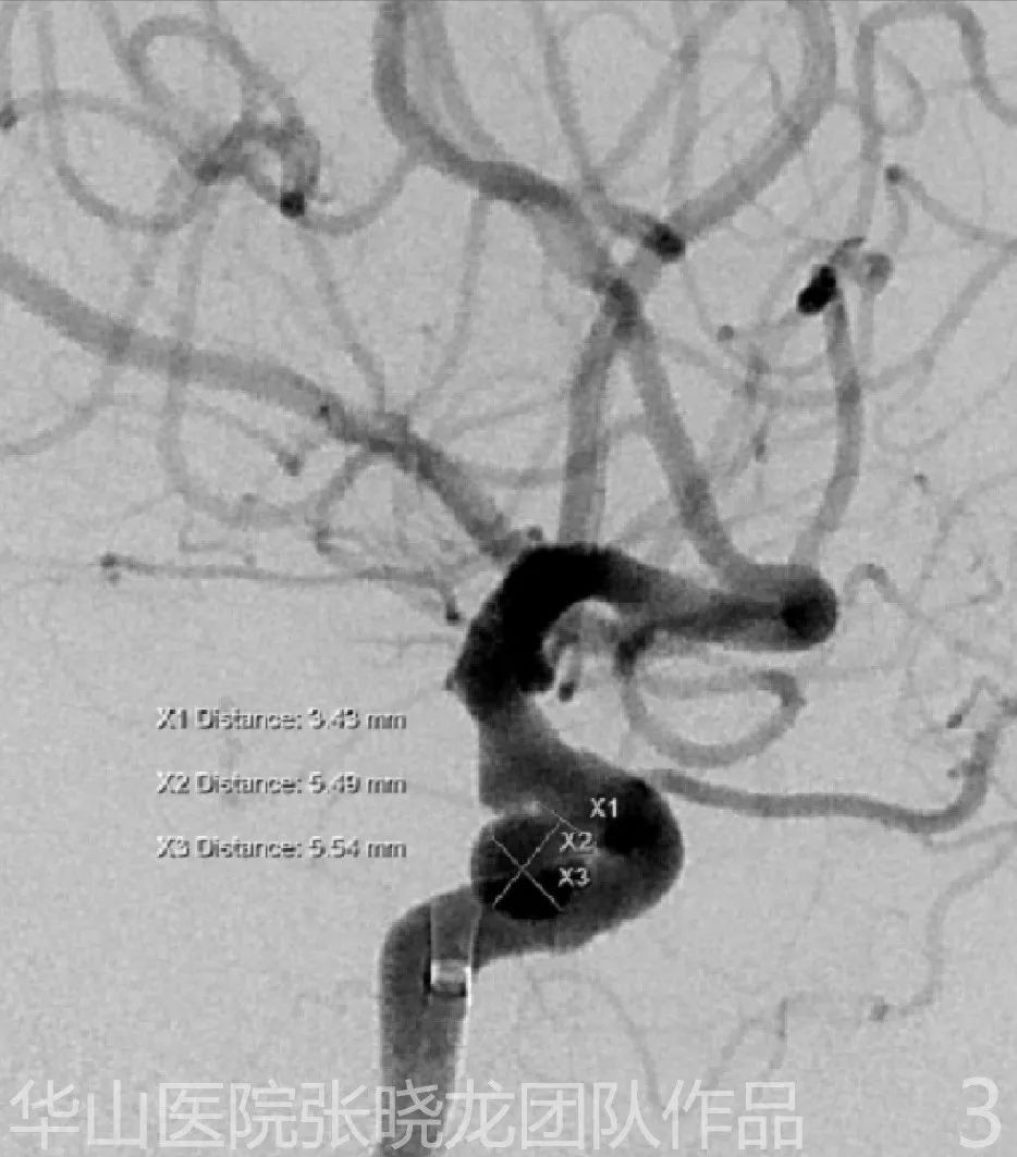
Figure 3. Maximal diameter: 5.5 mm. The working projection shows the necks of both aneurysms on a single image.
Video 3. 6F Envoy DA was advanced as far as possible via microcatheter and microwire. General heparinization. Echelon-10 microcatheter tip was shaped into a medial spiral curve.
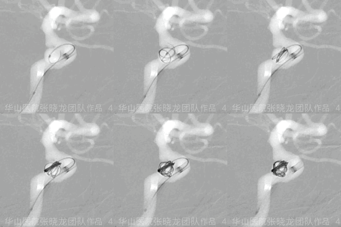
Figure 4 GIF. MicroPlex 6mm*20cm for framing.
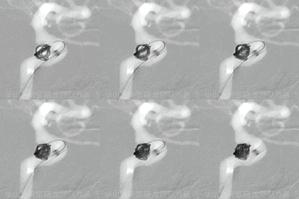
Figure 5 GIF. MicroPlex-10 5mm*15cm. MicroPlex-10 5mm*15cm。
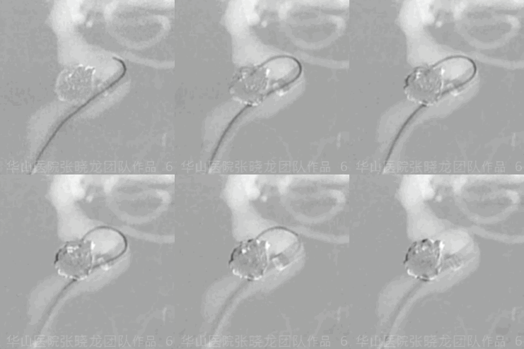
Figure 6 GIF. HydroCoil 4mm*8cm. The first Hydrocoil was difficult to insert. We kept the tension of the microcatheter and waited several seconds before inserting. HydroCoil 4mm*8cm。
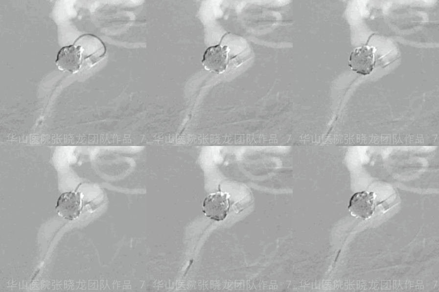
Figure 7 GIF. HydroCoil 3mm*8cm.
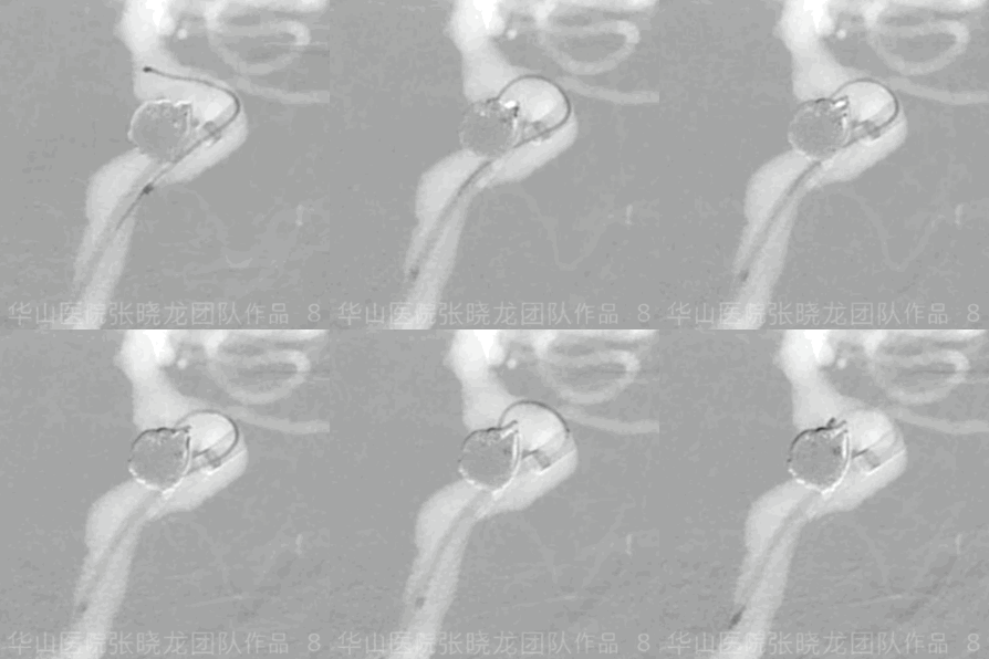
Figure 8 GIF. HydroCoil 2mm*3cm. The microcatheter was kicked out. We retrieved the microcatheter and re-navigated it into the inflow tract. The last coil densely packed the aneurysm neck. HydroCoil 2mm*3cm。
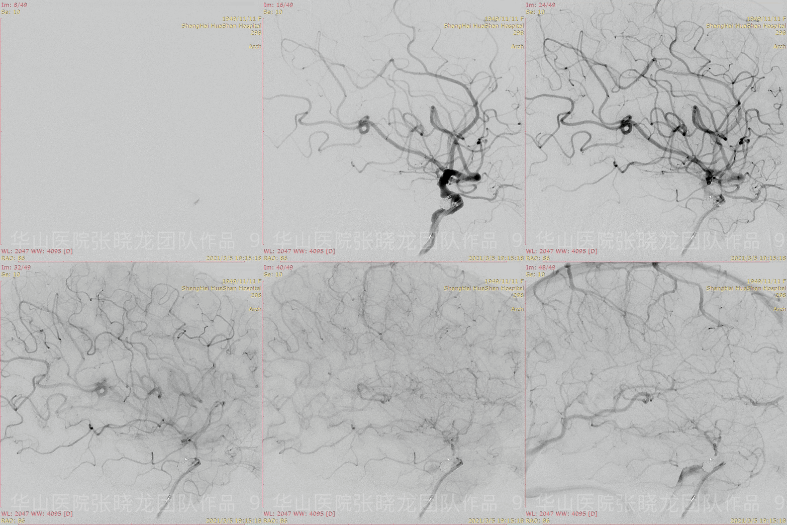
Figure 9 GIF. Working projection angiography shows the densely packing of the aneurysm with parent artery patent.
3
Post operation
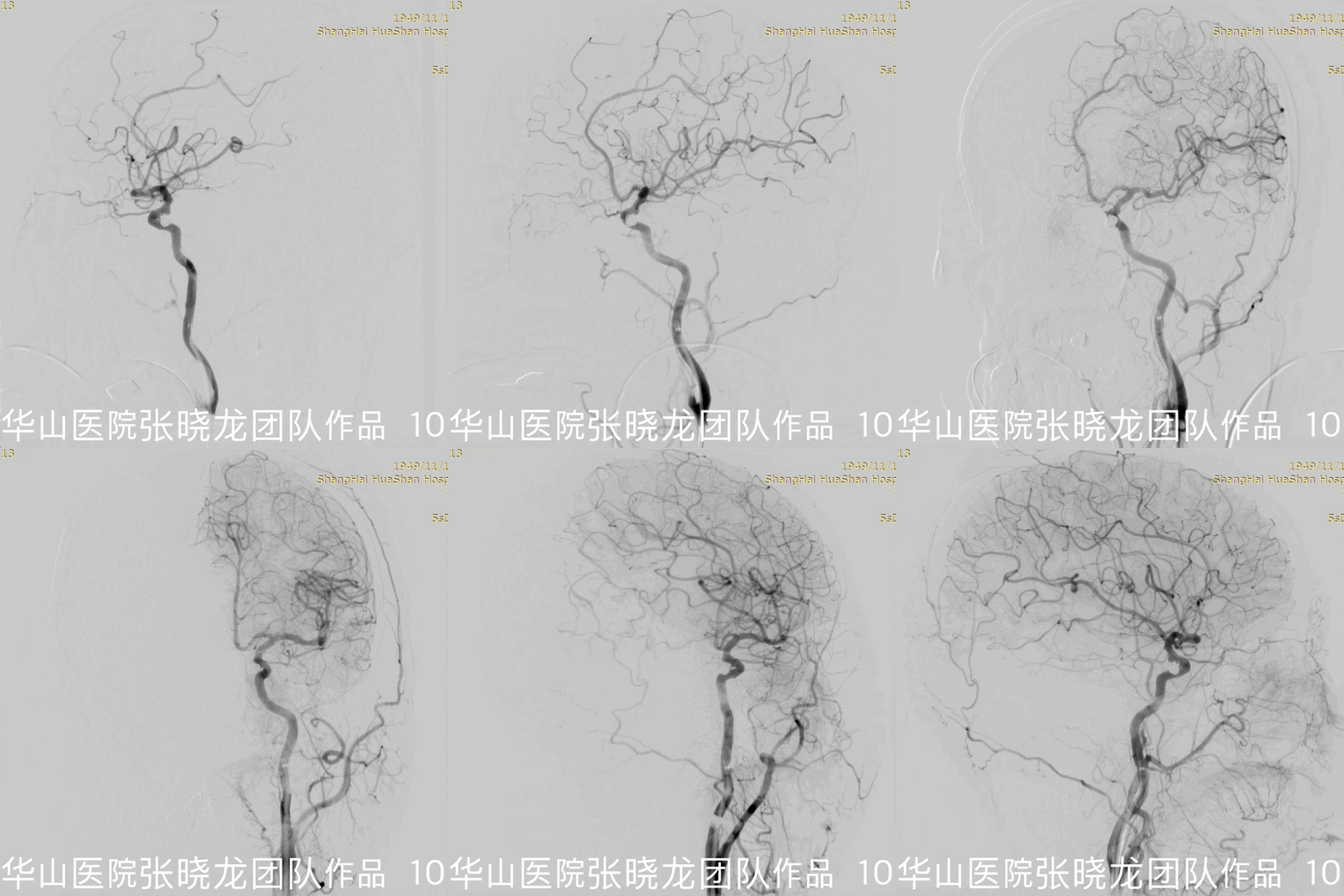
Figure 10 GIF. Post-operative rotational angiography shows the intact of intracranial vessels.
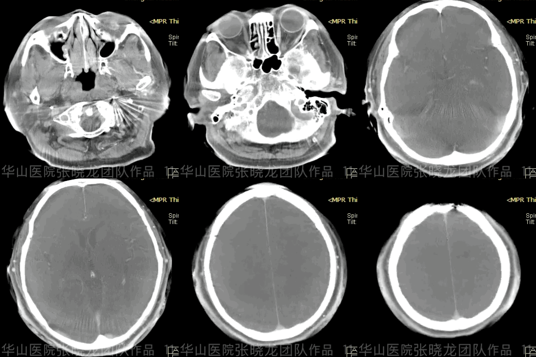
Figure 11 GIF. Post-operative Dyna CT shows no hemorrhage.
4
Summary
• Two tandem ophthalmic aneurysms in left ICA were revealed, one was regular-shaped and wide-necked indicating a low rupture risk, while the other is narrow-necked and therefore had a high rupture risk which could be treated with simple coiling.
• Large framing coil technique might decrease the recurrence rate while long term follow up DSA is necessary.
• Guiding catheter should be advanced across the petrosal curve as far as possible which can improve the maneuverability.
• The Hydrocoil is stiff and therefore difficult to insert. We kept the tension of the microcatheter and waited a few seconds to insert the third coil, a Hydrocoil 4*8.
• Two tiny carotid-ophthalmic aneurysms of the right ICA should be followed up.
