Published in February 23, 2023
Simple Coiling of an Ophthalmic Aneurysm
• 55 year-old female.
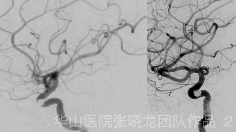
• Suffered from repeated headache for 2 years. DSA confirmed the right ophthalmic aneurysm 2 years ago. Video 1. DSA shows a right ophthalmic aneurysm.
• Simple coiling technique was attempted first.
• The microcatheter for stenting was spanpared in the parent artery. If simple coiling technique failed, stent assisted coiling would be selected.
Video 2. General heparinization. 6F Envoy. A Headway-21 microcatheter was advanced to the right MCA for stenting and protection. A Headway-17 microcatheter with spiral C curve was navigated to the aneurysm sac guided by looped microwire. 全身肝素化。视频 2. 6F Envoy导引导管。Video 3. Aneurysm size: 4.1mm*4.4mm, neck size:2.9mm. MicroPlex-10 5mm*15cm was inserted for framing.
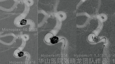
Figure 1. HyperSoft 4mm*8cm, 2mm*4cm, 1.5mm*4cm, 1.5mm*3cm were inserted into the aneurysm sac.
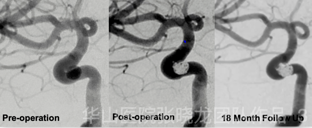
Figure 2 GIF. Post operative angiography shows densely packing of the aneurysm with the parent artery patent. No complication. GCS 15.
Figure 3. 18 month follow up angiography shows no relapse of the aneurysm. 1. In this case, choose stiff microcatheter & appropriate microcatheter shape both the stable framing of the first coil and the stability of the coiling microcatheter (headway 17) assure the method of simple coiling technique. Easier procedure, less complications.2. The first large coil formed a very stable frame which can spanvent recurrence.3. Next DSA follow up was suggested within 3-5 years4. No medication after follow up.
![article]()
Comments
Log into post your comments