
Case Review
History
• 63 y/o male.
• Suffering from weakness for 80 days and slow reaction and amnesia for 2 weeks.
• NE: (-).
• Medication: Valsartan, Aspirin, Atorvastatin.
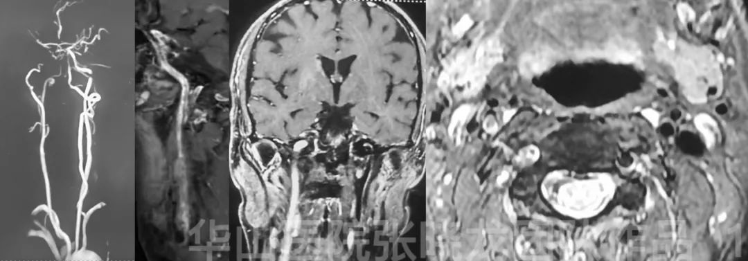
Figure 1. Multi-mode MRI shows the right ICA occlusion with enhanced thrombus.
Video 1. DSA shows the occlusion only involves the cervical segment of internal carotid artery.
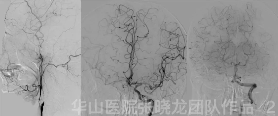
Figure 2. Collaterals via anterior communicating artery, external carotid artery and leptomeningeal are spansented.
1
Indication
• According to patient’s age, neurological symptoms, occlusion less than 3 month and the lesion limited in the cervical segment, recanalization of occluded ICA was performed.
2
Operation
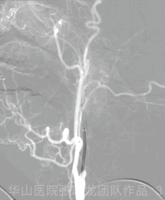
Figure 3 GIF. 6F Envoy. The guiding was navigated by Gateway 3mm*15mm and Transend 205 microwire to the cervical segment of the ICA. Balloon angioplasty was performed from distal to proximity of the ICA.
Figure 4 GIF. Angiography shows the recanalization of ICA with CCF formation.
Video 2. Two Precise 6mm*40mm stents were overlapping deployed from distal to proximal ICA.
Video 3. The initial segment of the occlusion was not well dilated.
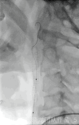
Figure 5 GIF. The stenotic initial segment was dilated by Litepac 4*30mm with 10-12 atm for 30s. HR decreased to 45bpm and returned to 65bpm after balloon dilatation.
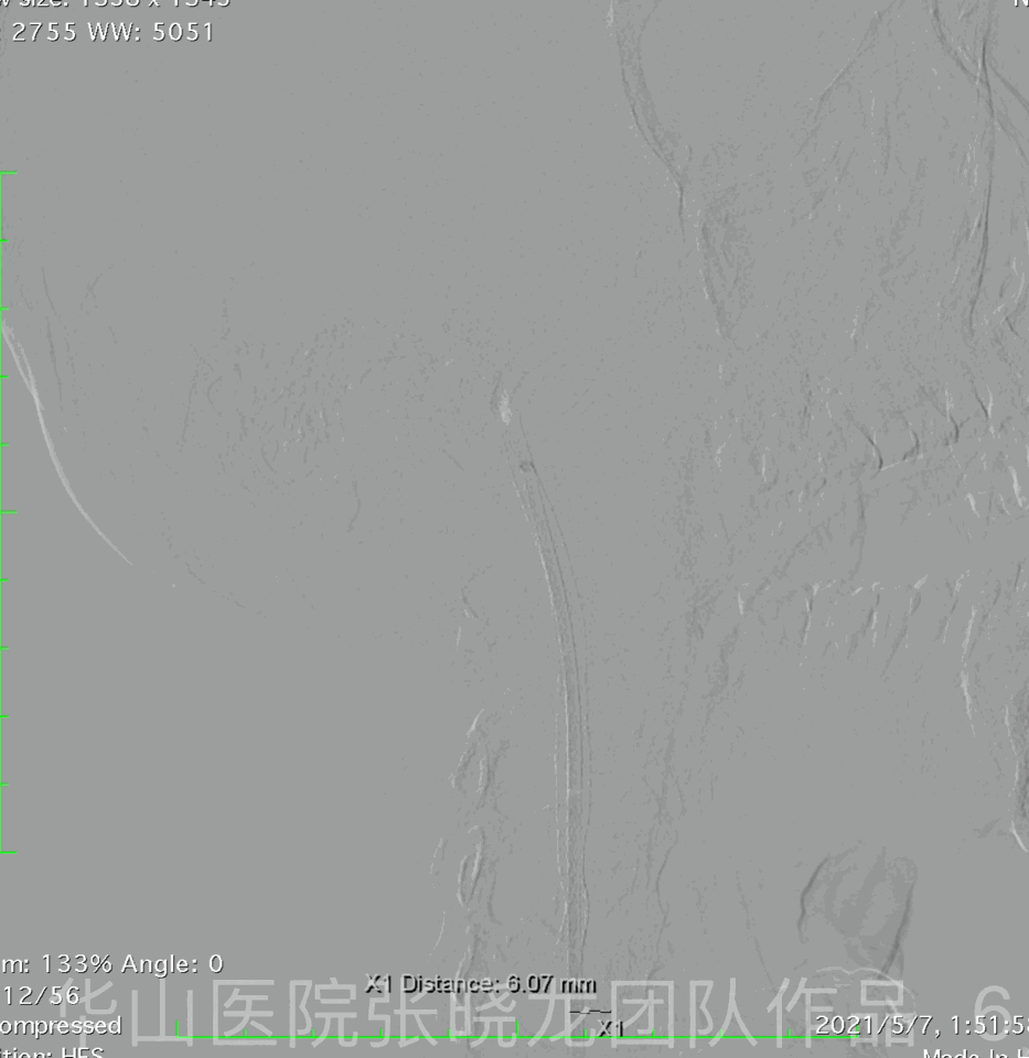
Figure 6 GIF. Angiography shows the occlusion of the petrosal and cavernous segment.
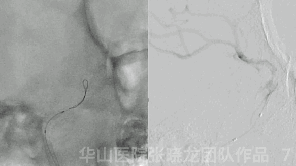
Figure 7 GIF. Guiding catheter was navigated to the distal cervical segment and Nimodipine 1ml was given. Gateway 3*15mm was advanced through the occluded segment and fluoroscopy confirms the system is in the real lumen of ICA. 导引导管到达颈段以远,给予尼莫地平1ml。
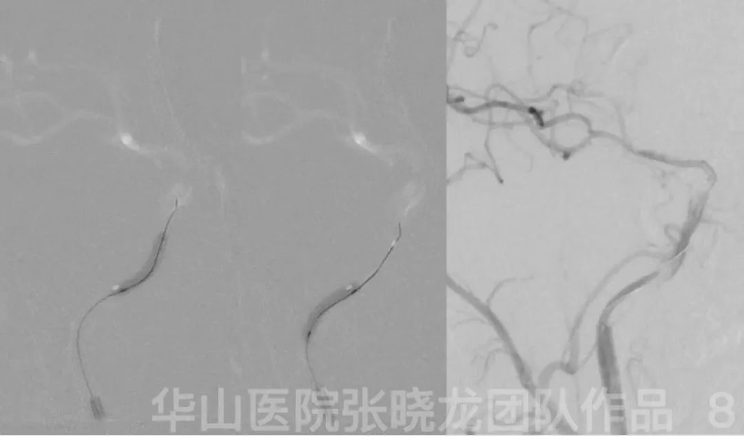
Figure 8. Balloon dilatation from the distal to the proximal of the ICA with 10-12 atm for about 40s.
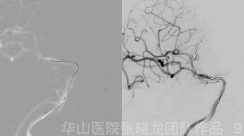
Figure 9 GIF. Re-navigation of Gateway 3*15mm. Angiography shows the recanalization of the occusion.
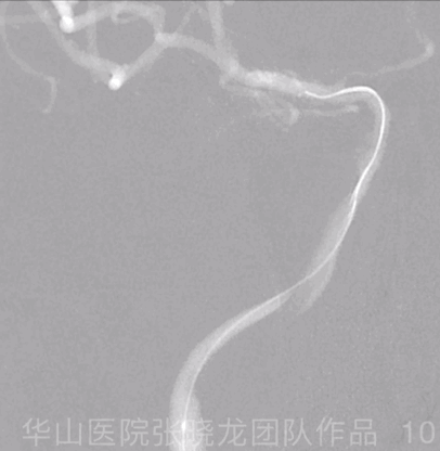
Figure 10 GIF. Prowler Plus microcatheter was advanced to the M1.
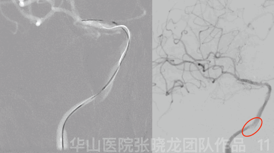
Figure 11 GIF. Enterprise 4.5*37mm was deployed from distal to proximal segment of the ICA. Angiography shows the proximal dissection.
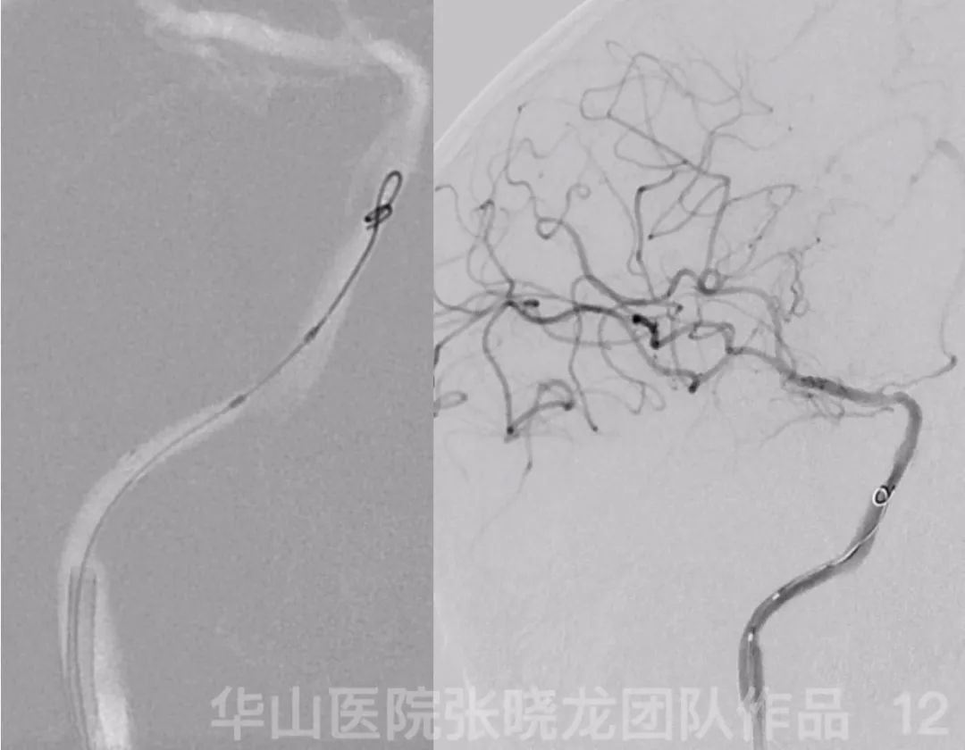
Figure 12. The dissection was well dilated by Gateway 4*9mm with 4atm for 40s.
Video 2. Post operation angiography shows satisfied dilation of the stenosis with mild stenosis left. Tirofiban 16ml. No general heparinization.
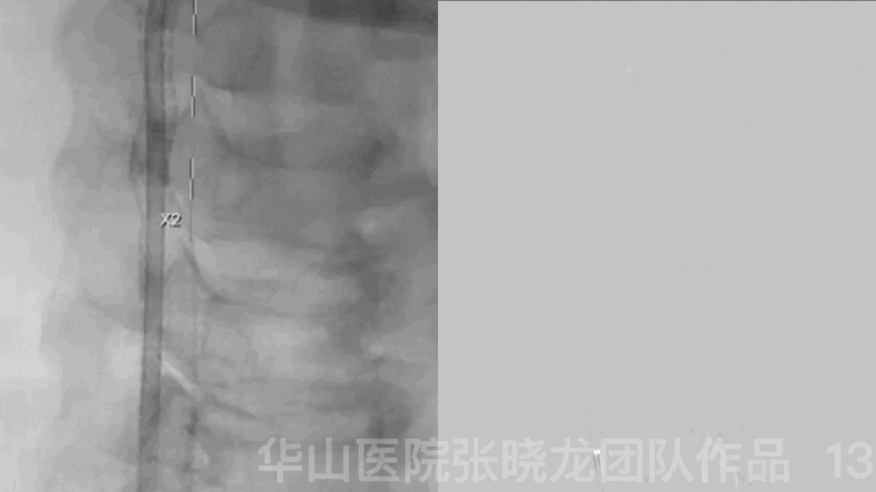
Figure 13 GIF. Third Precise 6mm*40mm was deployed in the initial ICA followed by Litepac 5*30mm with 4atm for 10s.
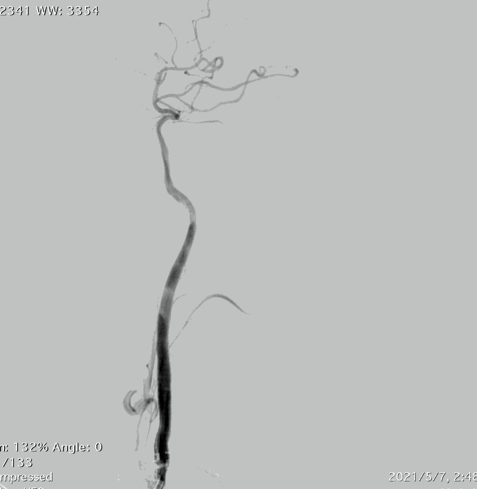
Figure 14 GIF. Post-operative angiography shows the complete recanalization of ICA.
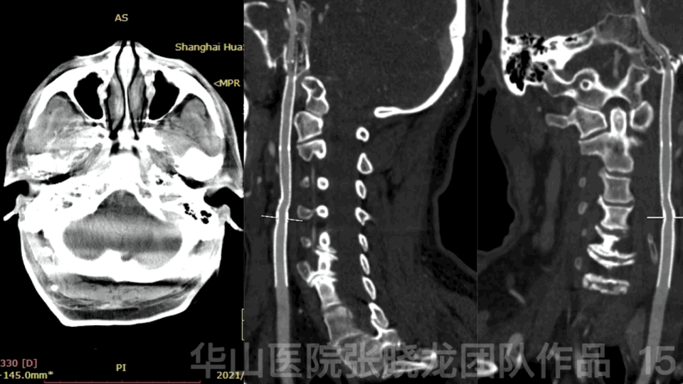
Figure 15 GIF. Dyna CT shows no hemorrhage.
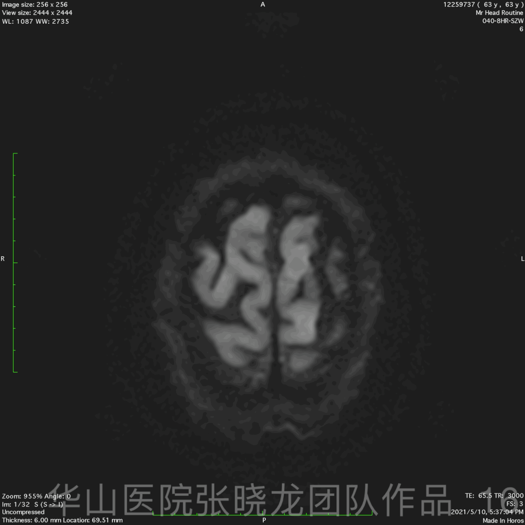
Figure 16 GIF. Post-operative DWI shows no acute infarction.
3
Post operation
• No new neurologic deficit.
• Control SPB 100-130mmHg
• Dual antiplatelet therapy 24 hours later
4
Summary
• According to patient’s age, neurological symptoms, occlusion less than 3 month and the lesion limited in the cervical segment, recanalization of occluded ICA was performed.
• 6F Envoy with Gateway balloon can supply better support to across the occluded segment.
• Two Carotid stents were deployed to avoid the thrombus immigration and vessel reconstruction.
• It is acceptable that the microcatheter was navigated to the real lumen after passing the dissection at the external cranial segment of the ICA.