
Our case
History
• 69 y/o female
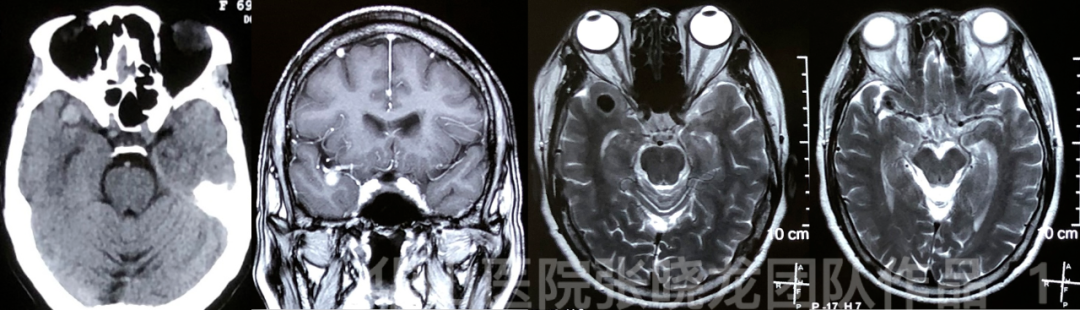
Figure 1. MR and CT shows a right MCA aneurysm.

Figure 2. DSA confirms the diagnosis of the right MCA aneurysm. Proximal ICA vasospasm indicates fragility of the vessel walls.
1
Strategy
• Stent-assisted coiling
2
Operation
Video 2. As the cervical ICA was very fragile, a 70cm long sheath was advanced to the CCA, while a Navien 105cm 072 catheter was navigated to the cavernous segment. Nimodipine 1ml was administered before advancing guiding catheter.
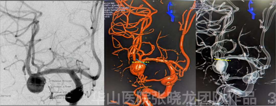
Figure 3. Measurement.

Figure 4 GIF. Prowler Plus microcatheter with C curved tip was advanced to the inferior branch of the right MCA with the guide of Synchro-2 soft microwire with S curved tip.

Figure 5. Superselective angiography by microcatheter shows the microcatheter is placed in the true lumen of vessel.
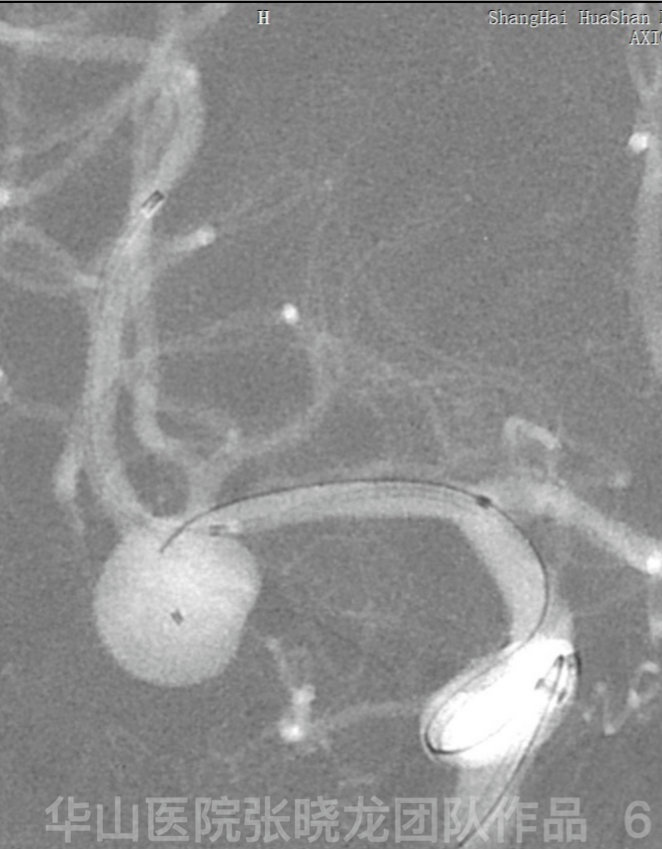
Figure 6. Echelon 10 45° microcatheter with C curved tip was advanced to the aneurysm sac. General heparinization.
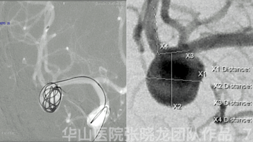
Figure 7 GIF. The width was 10.18mm, a MicroPlex-18 13mm*32cm framing coil was selected for better stability.
Video 3. Solitaire 4mm*20cm stent was deployed in the inferior branch of the right MCA.
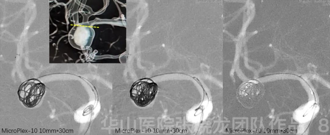
Figure 8. Then the aneurysm sac was packed with MicroPlex-10 coils to ensure full deployment of the stent. Five MicroPlex-10 10mm*30cm coils were packed within the frame, and the superior branch was spanserved well.
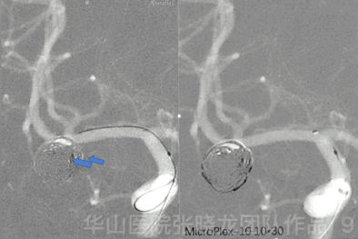
Figure 9 GIF. To fill the outflow tract.
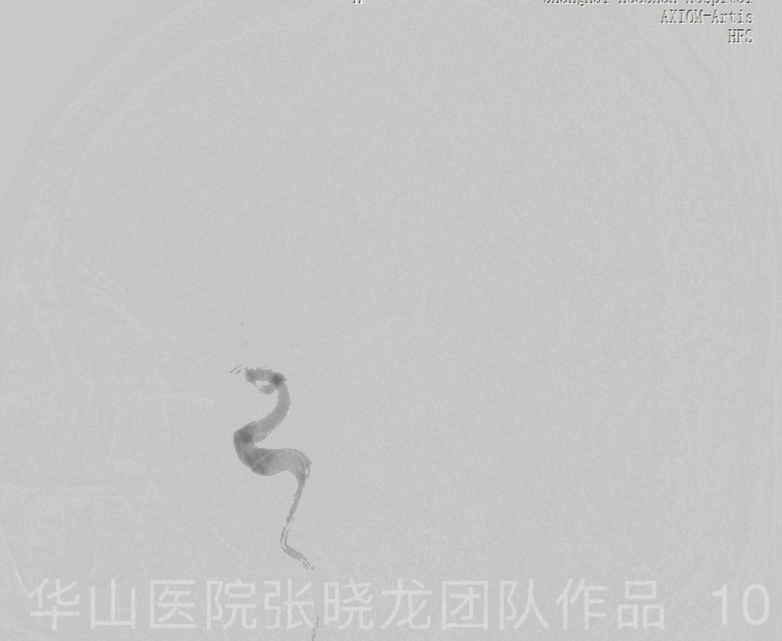
Figure 10 GIF. Angiography shows the remnant of the inflow tract.
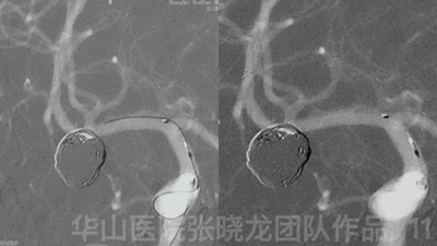
Figure 11 GIF. MicroPlex-10 5mm*15cm (x4) coils were inserted for packing the inflow tract of the aneurysm.
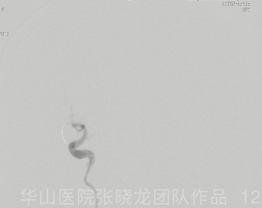
Figure 12 GIF. Post-operative angiography shows the densely packing of the aneurysm with the parent artery patent. Tirofiban (Xinweining) 12ml was administrated.
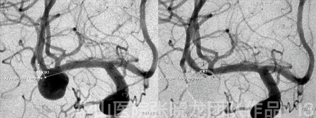
Figure 13. Parent artery angle increases from 103.1° to 136.3°.
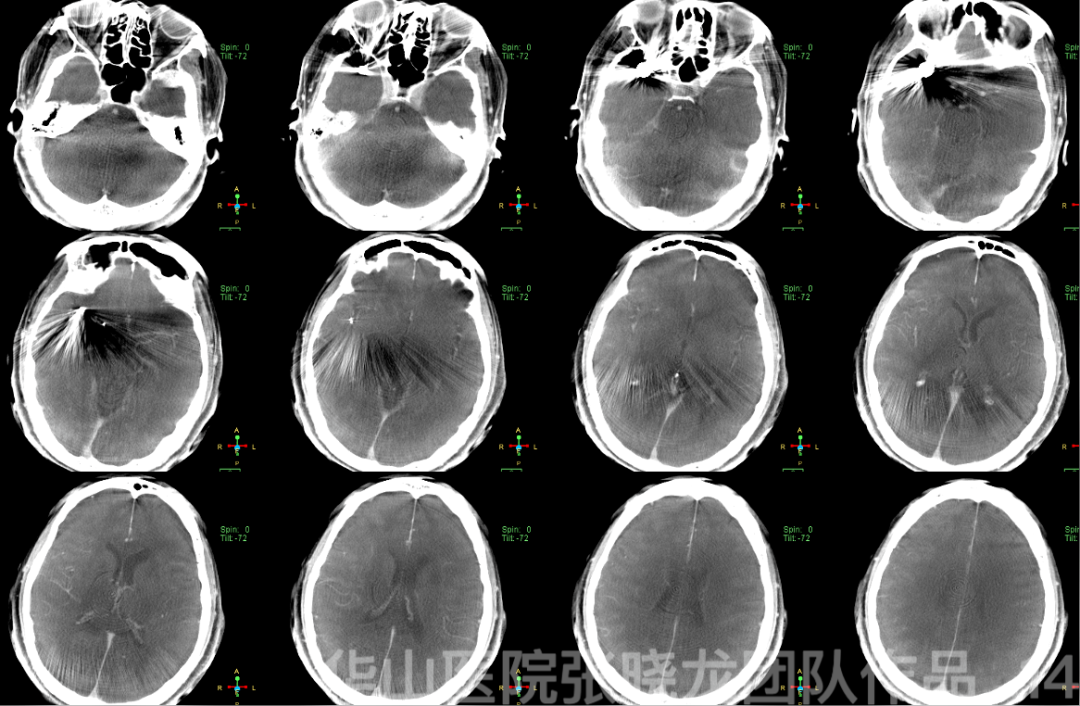
Figure 14. Post-operative CT shows no hemorrhage or large acute infarction.
3
Post operation
• No complication. NS (-)
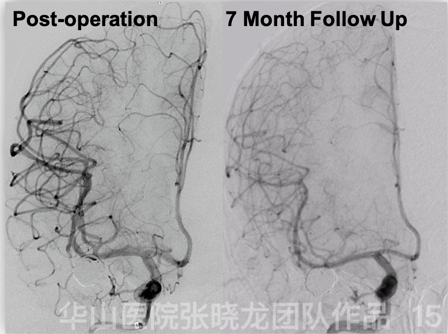
Figure 15. Seven month follow up angiography shows no relapse of the aneurysm. The patient was symptom free.
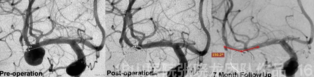
Figure 16. The change of aneurysm angle from span-operation to follow up: 103.1°→ 136.3°→150.2°.
4
Summary
• MCA bifurcation aneurysm size: 9.7mm*10.2mm; Neck: 6.02mm;