Case Review
History
1
Strategy
• Bilateral MCA aneurysms need to be treated at the first stage.
2
Left MCA aneurysm
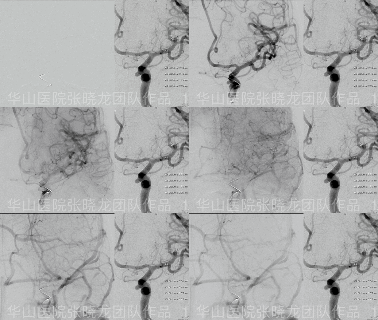
Figure 1 GIF. 6F Envoy DA. General heparinization. 6F Envoy DA.
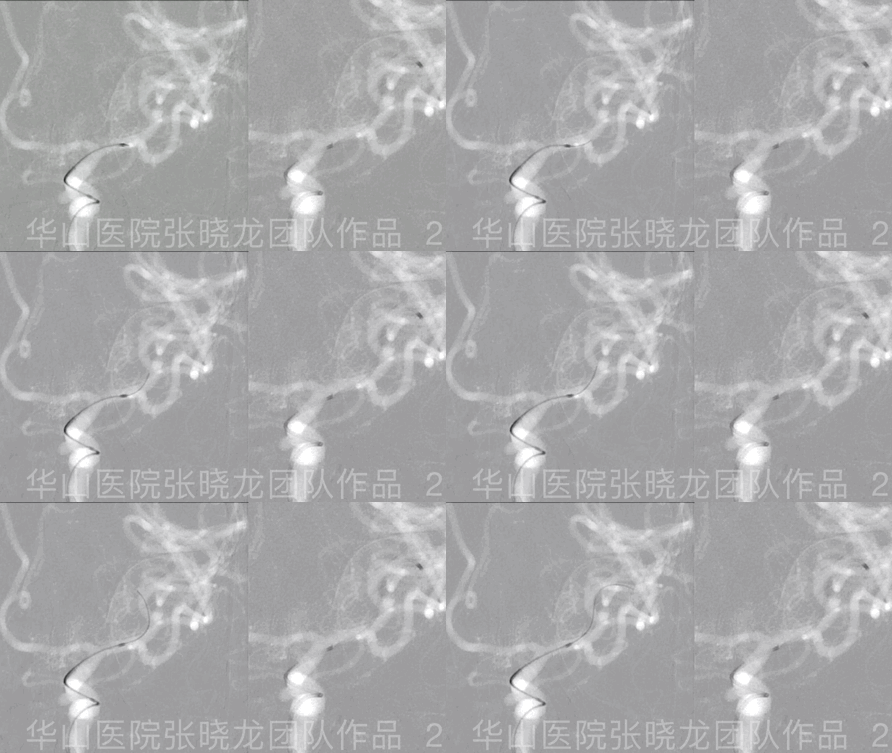
Figure 2 GIF. A Prowler Plus microcatheter with C shaped tip was navigated to the M2 segment of the superior MCA branch guided by Synchro II microwire.
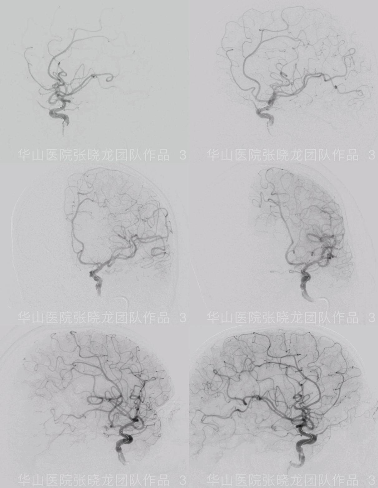
Figure 3 GIF. Due to remodling of the parent artery after stenting,repeated rotational angiography was performed to find a new working projection for coiling.
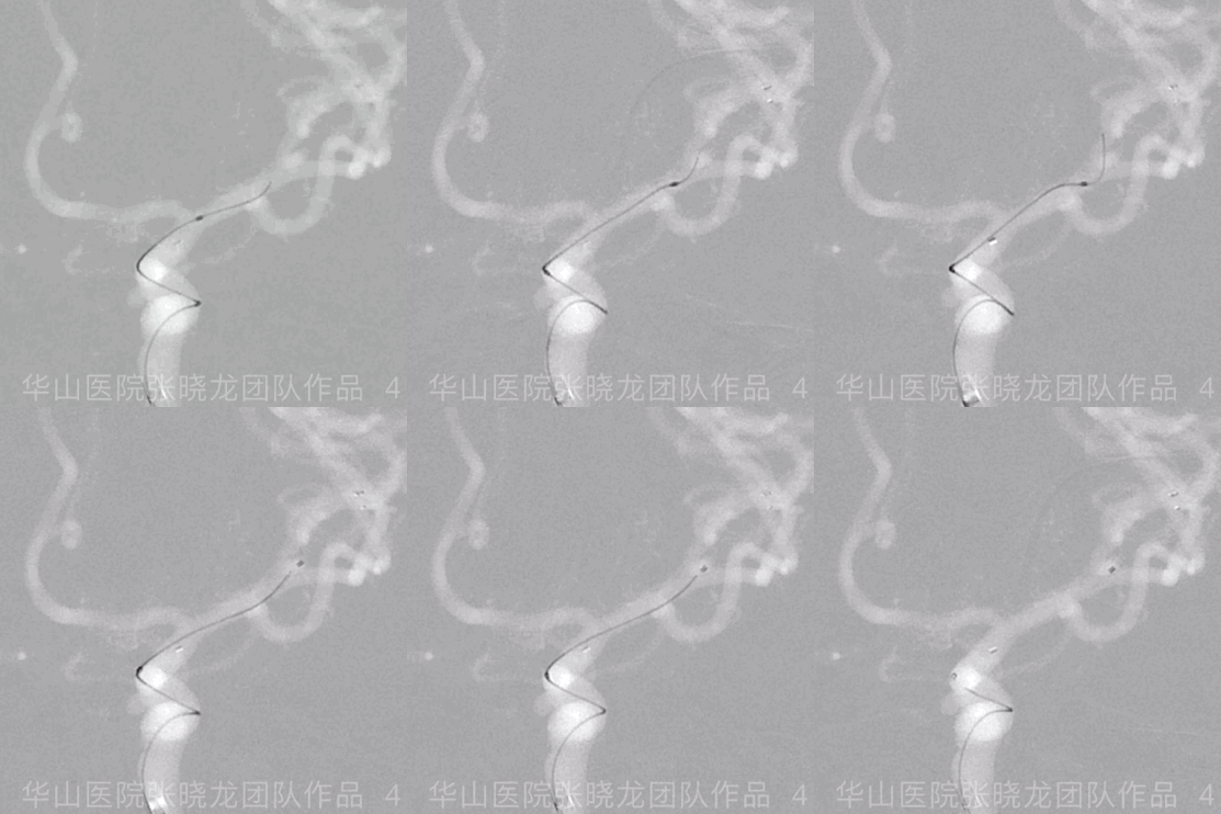
Figure 4 GIF. Echelon-10 with straight tip was advanced to the aneurysm sac.
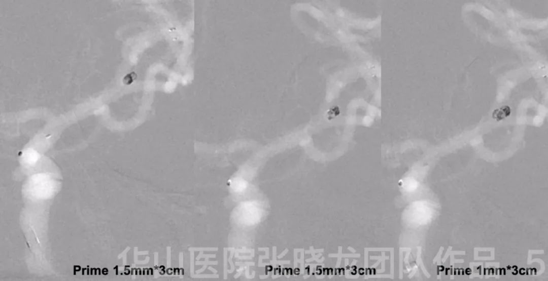
Figure 5. Working Projection I. Prime 1.5mm*3cm (2) and Prime 1mm*3cm coils were inserted into the aneurysmal sac.
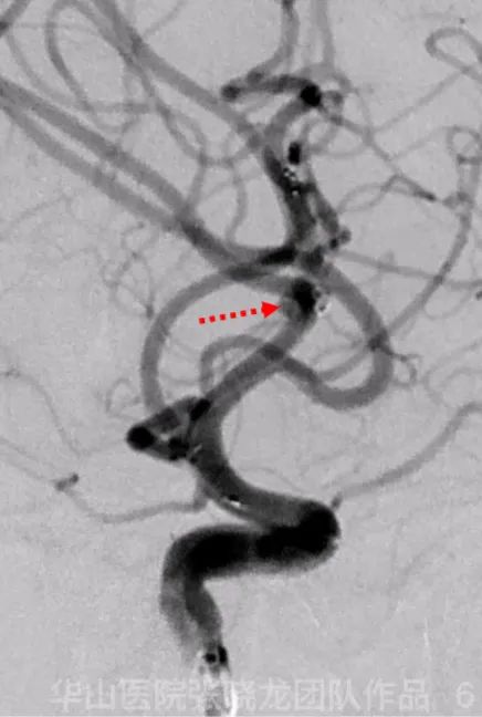
Figure 6. Working Projection II shows the other daughter sac.
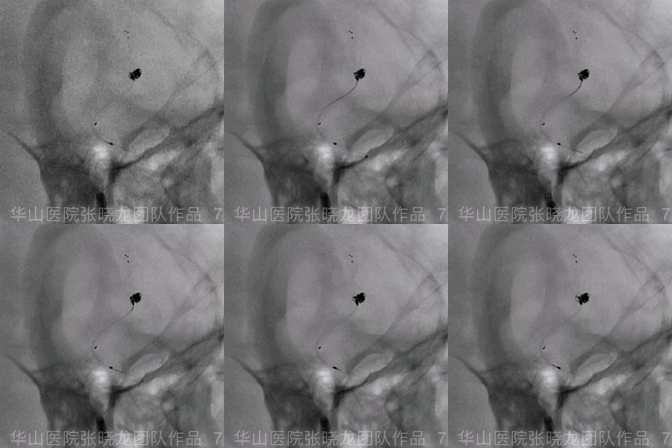
Figure 7 GIF. The tip of the coiling microcatheter was adjusted.
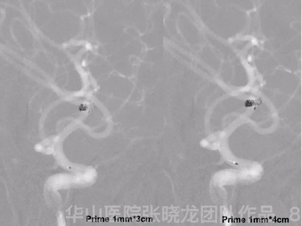
Figure 8. Prime 1mm*3cm and Prime 1mm*4cm coils were inserted into the aneurysm sac.
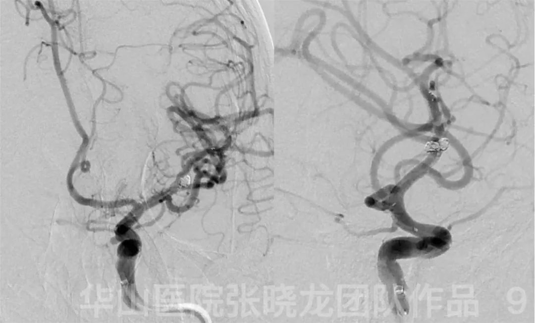
Figure 9. No intra-operative complication occurred. Tirofiban 15ml was administrated via the guiding catheter.
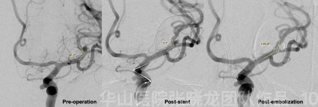
Figure 10. After the treatment, the parent artery angle increased from 97.2 degrees to 140.0 degrees.
3
Right MCA aneurysm
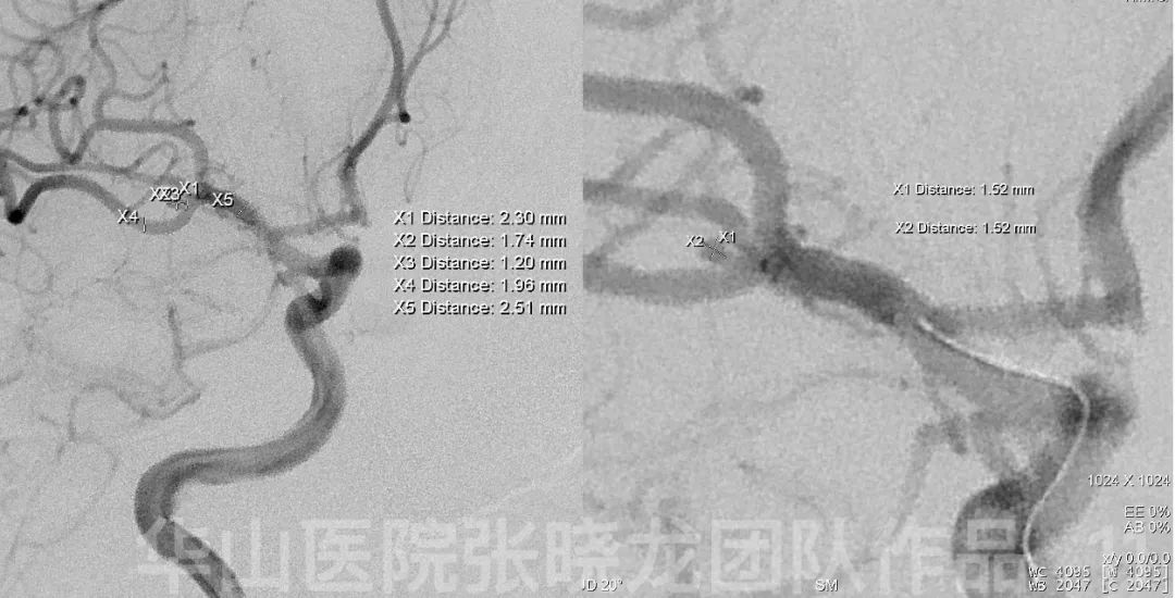
Figure 11. Measurement of the right MCA aneurysm.

Figure 12. 6F Envoy DA. A Prowler Plus microcatheter with C shaped tip was navigated to the M2 segment of the right inferior MCA branch guided by a Synchro II microwire.
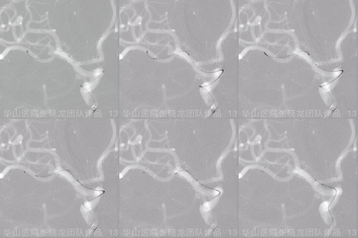
Figure 13 GIF. Solitaire 4*20mm was deployed covering aneurysm neck and spanserving the inferior branch of the right MCA.
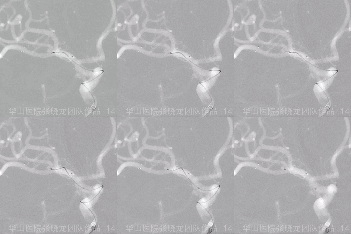
Figure 14 GIF. An Echelon-10 microcatheter with a straight tip was advanced to the aneurysm neck.
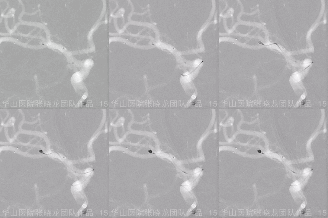
Figure 15 GIF. A Perdenser 1.5mm*3cm coil was inserted into the aneurysm sac.
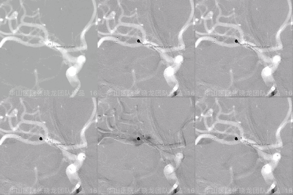
Figure 16 GIF. A Prime 1mm*2cm coil was failed to be inserted. Fluoroscopy shows the leak of contrast medium indicating aneurysmal rupture.
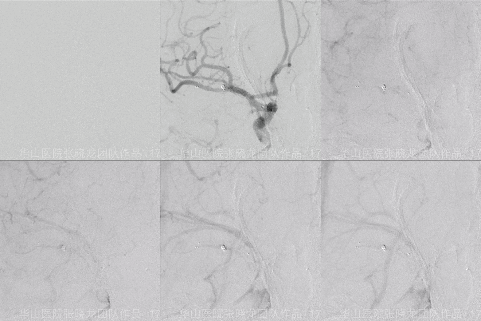
Figure 17 GIF. No more bleeding of the rupture site is observed.
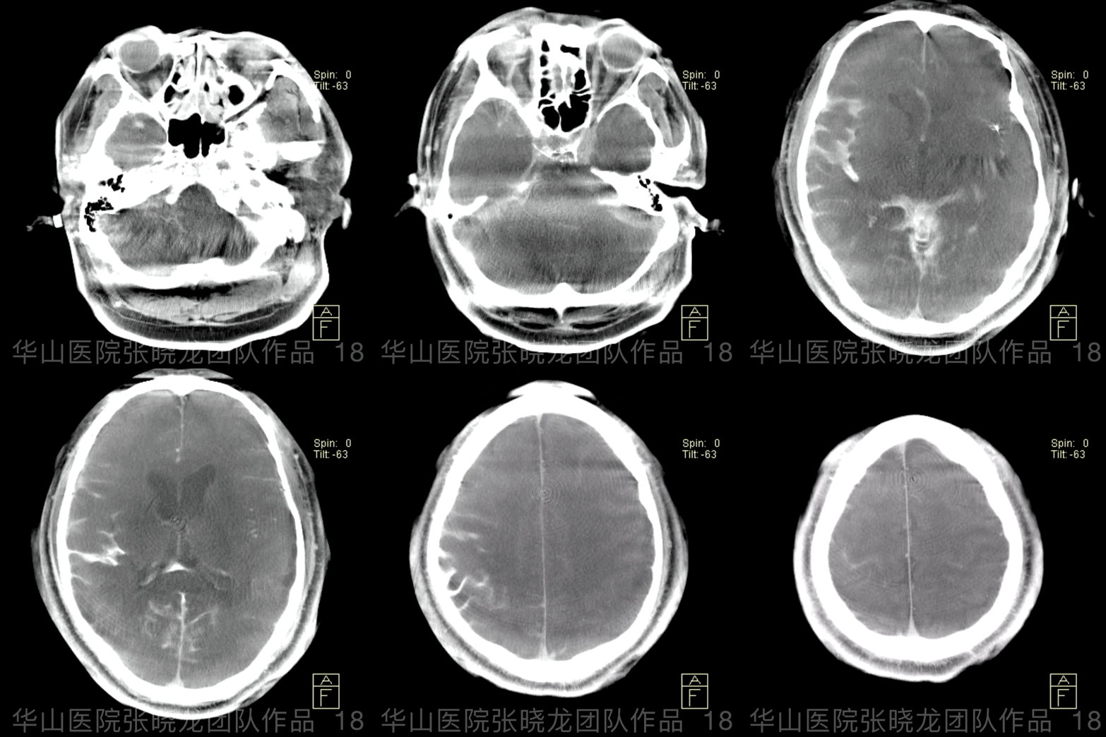
Figure 18 GIF. Emergency Dyna CT confirms SAH.
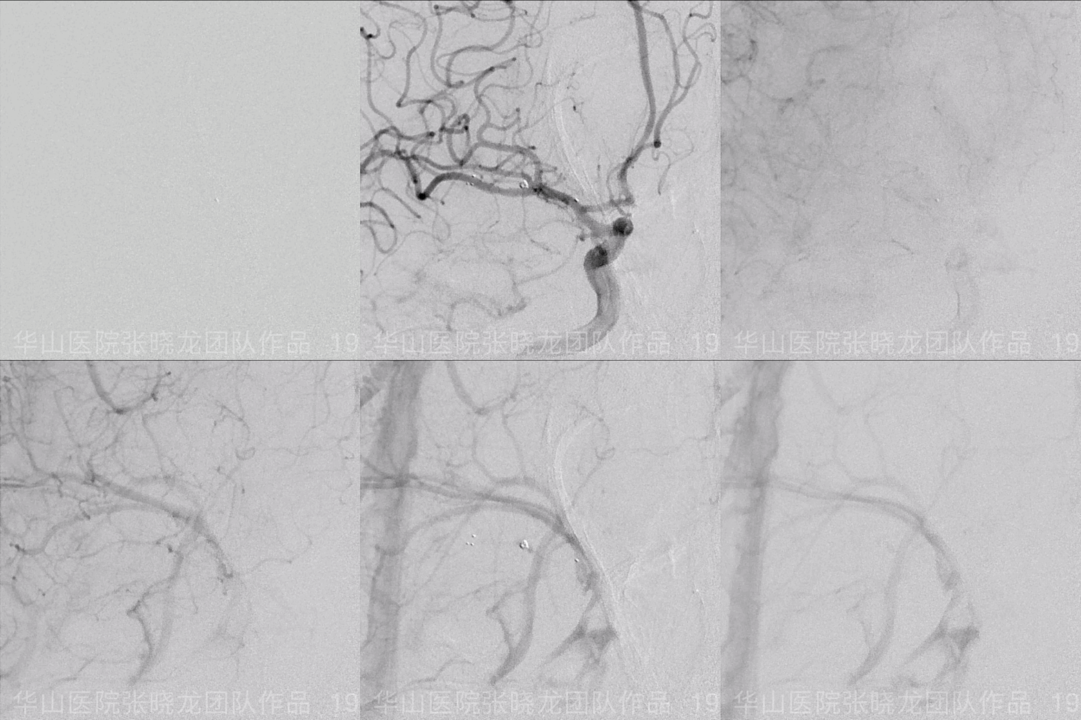
Figure 19 GIF. Angiography shows no bleeding after 20 mins waiting.
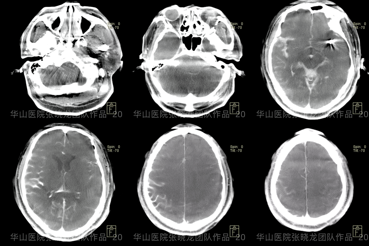
Figure 20 GIF. Second Dyna CT shows no increasing of bleeding volume or hydrocephalus.
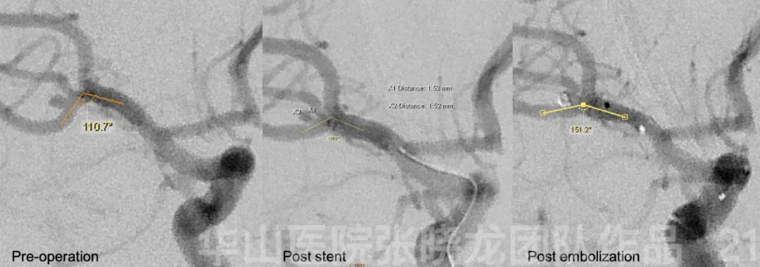
Figure 21. The parent artery angle increased from 110.7 degrees to 151.2 degrees.
4
Post operation
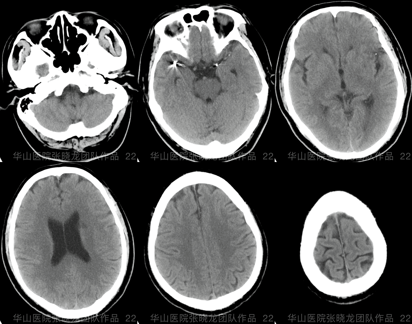
• PE: fever 38.3, aphasia, right limbs muscle strength were grade 4. And 3 days later, all symptoms were recovered.
• Medication: In 48h, Furusemide 20mg, Mannitol 100ml, Serum Albumin 10g q12h, after 48h, and Aspirin 100mg qd.
5
Discharge
• No fever or neurologic deficit.
• Medication:Aspirin 100mg qd, Clopidogrel 75mg qd,Irbesartan 150mg qd.
6
Summary
• Comspanss the right ICA
7. Increased parent artery angle after the treatment may lower the risk of aneurysm relapse.
