
Case Review
History
• 53 y/o, male
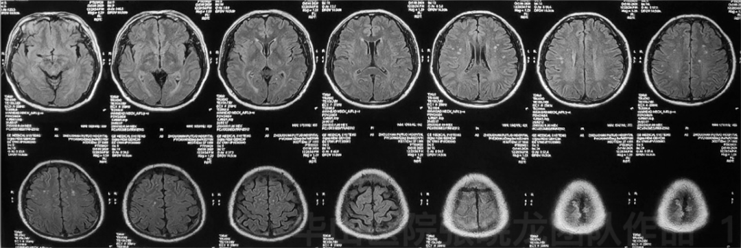
Figure 1. T2 Flair shows scattered small infarctions.
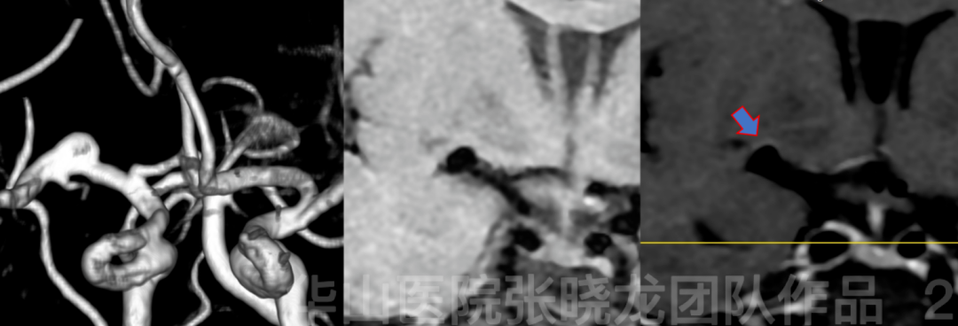
Figure 2. Multi-model MRI shows a right M1 dissecting aneurysm with partial enhanced aneurysmal wall.
Video 1. Angiography confirms a right MCA dissecting aneurysm incorporating lenticular arteries (red arrow).
1
Strategy
SAC (Stent-assisted coiling)-Decrease recurrence rate
2
Operation
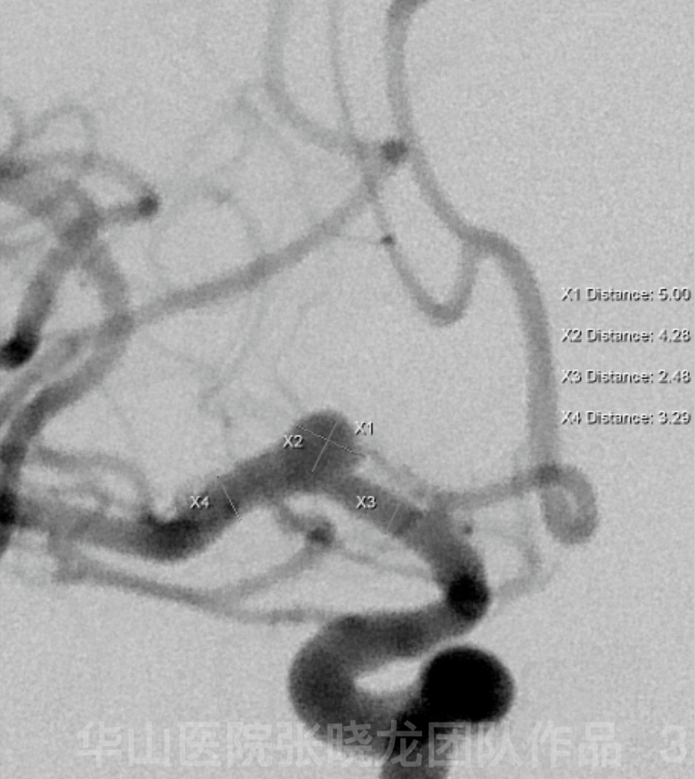
Figure 3. Measurement.
Video 2. General heparinization.6F guiding catheter. A Prowler plus microcatheter was advanced to the M2 branch guided by a Synchro-2 microwire. An Echelon-10 microcatheter with a straight tip was navigated to the aneurysmal sac. Microplex-10 4mm*10cm coil was selected for framing which was unsatisfactory.
Video 3. A Solitaire 4*20mm stent was deployed via the Prowler Plus microcatheter.
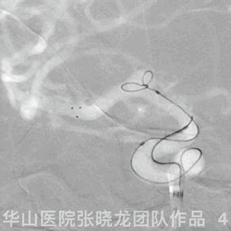
Figure 4 GIF. A successful coil framing was made by the Microplex-10 4mm*10cm after stent deployment. At the same time, the tip of the coiling microcatheter was pushed out of the aneurysmal sac.

Figure 5 GIF. The tip of the coiling microcatheter was renavigated to the aneurysmal sac guided by a microwire.
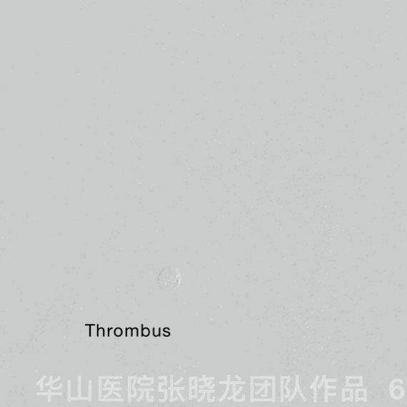
Figure 6 GIF. Six coils were inserted into the aneurysmal sac. Hypersoft 3mm*8cm (1), Hypersoft 1.5mm*4cm (2), Hypersoft 2mm*3cm (1) ,Target 360 2mm*3cm (1), Target 360 2mm*4cm (1). But post-coiling angiography shows thrombosis in the parent artery and suspected hemorrhage.

Figure 7 GIF. Repeated angiography shows no hemorrhage.
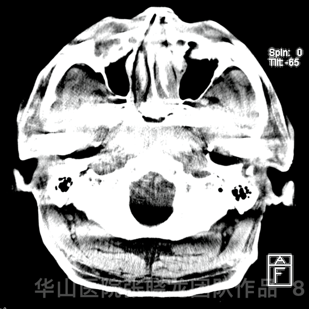
Figure 8 GIF. Dyna CT also confirms no hemorrhage.

Figure 9 GIF. After given Tirofiban 16ml and Nimodipine 1ml via the guiding catheter, the angiography shows complete dissolution of the thrombus and patent lenticular arteries arising from the neck region of the aneurysm.
3
Post-operation
• No neurologic deficit.
• Medications: Clopidogrel 75mg qd for three months, Aspirin 100mg qd until next follow up, anti-hypertensive medicine.
4
Follow up

Figure 10 GIF. Seven-month follow up angiography shows densely packing of the aneurysm with parent artery patent.
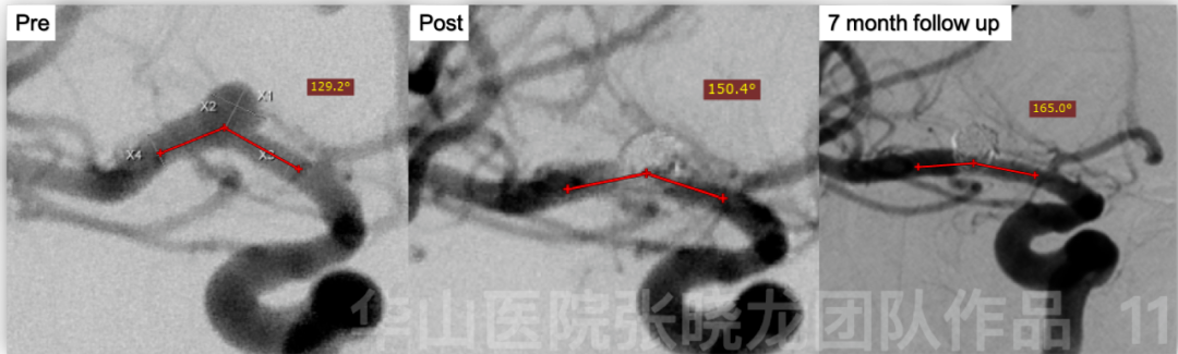
Figure 11. Compared with span and post treatment angiography, follow up angiography shows the parent artery angle increases from 129.2 degrees to 165.0 degrees.
4
Summary
• Indications: aneurysm wall enhancement and ischemic symptoms