
Case Review
History
Video 1. Angiography shows two ophthalmic artery aneurysms with ipsilateral ICA dissection. One of the ophthalmic aneurysm is on the course of oculomotor nerve.
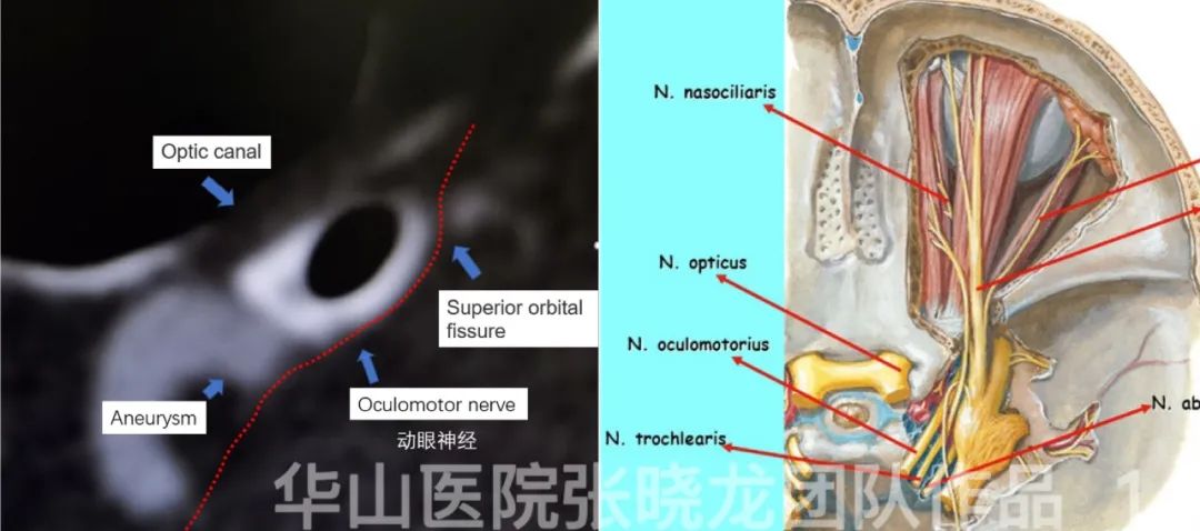
Figure 1. Ptosis was due to the left oculomotor nerve comspanssed by the aneurysm.
1
Strategy
Two aneurysms located at the ophthalmic segment of the left ICA.
Stent-assisted coiling – it may be difficult to embolize the second aneurysm after stent deployment. FD stent can simplify the procedure.
2.The ICA dissection can be treated with carotid stent implantation. During the procedure, the guiding catheter should be first navigated into the cavernous segment of the ICA for FD deployment. Then retrieve the guiding catheter and deploy the carotid stent.
2
Operation
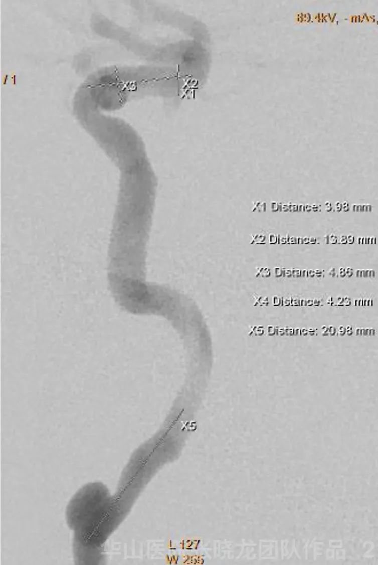
Figure 2. General Heparinization was administrated at the beginning of the operation.
Figure 3 GIF. 6F 90cm sheath. 6F 115cm Navien was placed at the cavernous segment of the left ICA.
Figure 4 GIF. Marksman microcatheter was navigated to the left M1 guided by Synchro II standard microwire.
Video 2. Pipeline 4.0*20mm was deployed covering two ophthalmic artery aneurysms.
Figure 5 GIF. Precise 6*40mm stent was deployed for remodeling the dissecting segment of the left ICA.
Figure 6 GIF. Post-operation angiography shows the contrast medium retention in the aneurysm sac and the dissection.
Figure 7 GIF. 3D reconstruction shows the well-deployment of the Pipeline stent.
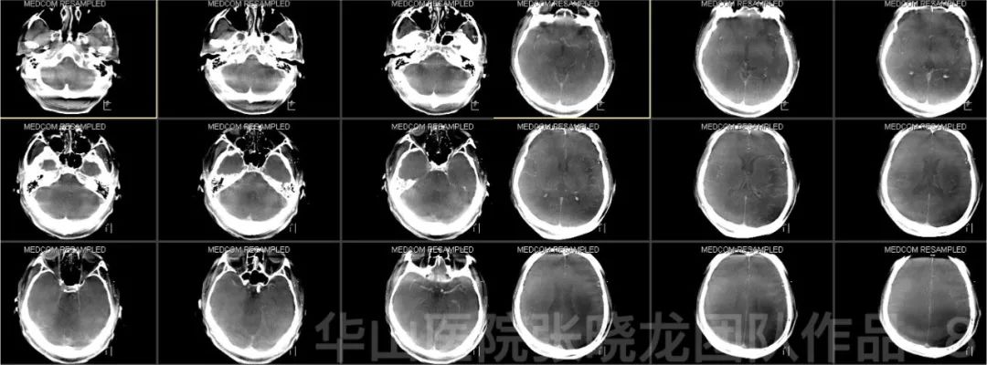
Figure 8. Post-operative Dyna CT shows no intracranial hemorrhage or infraction.
3
Post-operation
• No new neurologic deficit.
4
Follow up
3-month follow up
• Dual antiplatelet therapy was used for one month.
• Therapy was stopped by the patient himself.

Figure 9 GIF. 3-month follow up angiography shows the complete occlusion of two ophthalmic artery aneurysms with patent parent artery and mild stenosis proximal to the dissection.
5
Summary
• In the case with two ophthalmic artery aneurysms, flow divert treatment is recommended for simplifying the procedure, which is also beneficial to oculomotor nerve palsy recovery.