Case Review
History
Figure 1. CT scan demonstrates left MCA AN was embolized. Left watershed had malacia and chronic hemorrhagic foci.
Figure 2. MRI DWI sequence reveals no acute infarction.
1
Strategy
2
Operation
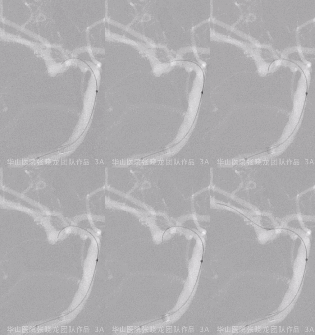
Figure 3A. Headway 21 microcatheter was navigated into position.
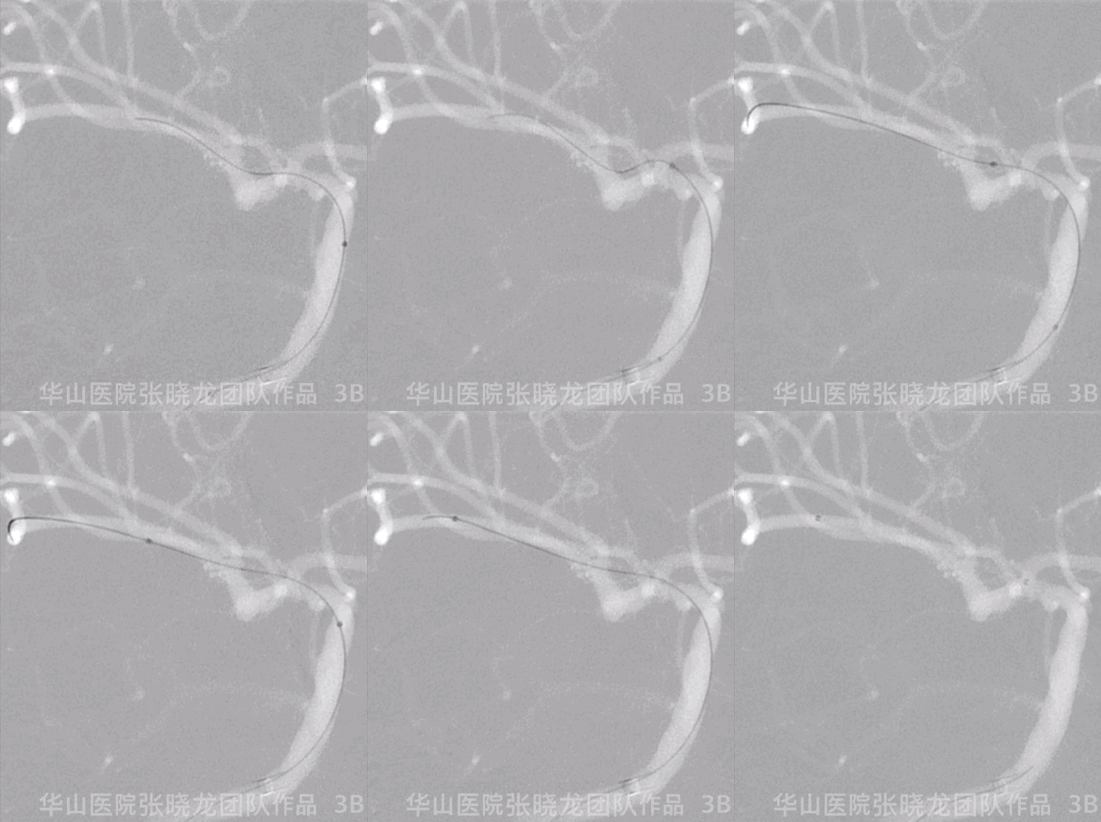
Figure 3B. Headway 21 microcatheter was navigated into position.

Figure 4 GIF. Right MCA M1 segment was straightened.
Figure 5. AN Measurements.
Video 3. Headway-17 microcatheter with C curve was navigated into the aneurysm sac.

Figure 6 GIF. 1st coil Hypersoft 3mm*6cm.
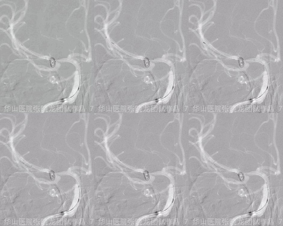
Figure 7 GIF. Solitaire 4mm*20mm was placed for protecting superior branch of right MCA.
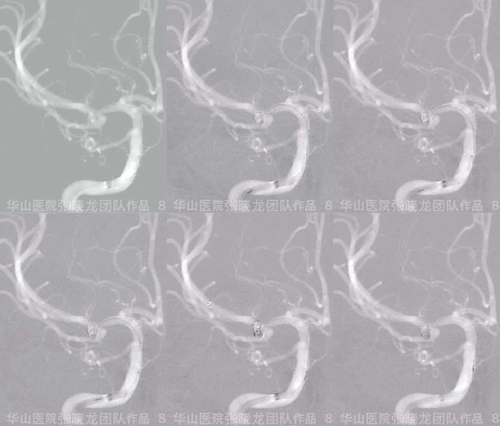
Figure 8 GIF. Hypersoft 2mm*4cm.
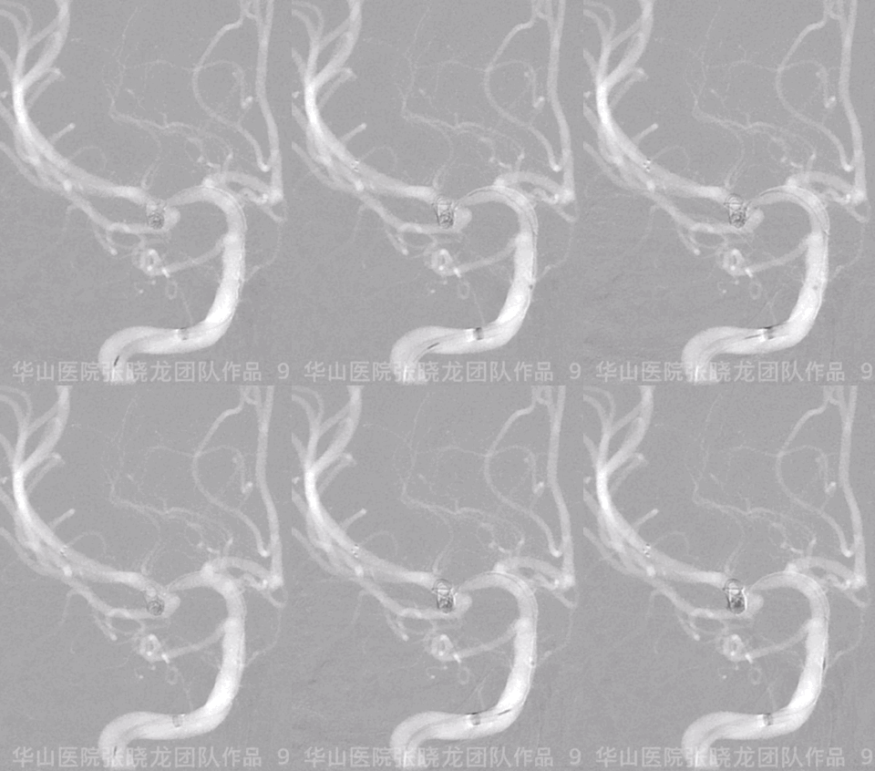
Figure 9 GIF. Hypersoft 2mm*4cm.
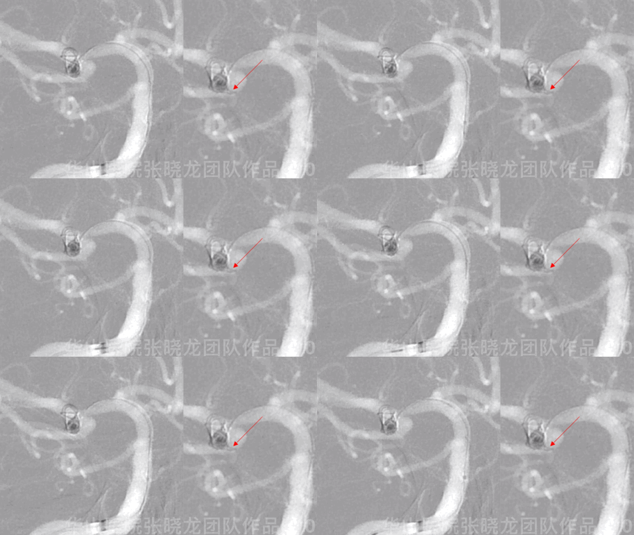
Figure 10 GIF. The coil (Hypersoft 2mm*4cm) punctured the aneurysm sac.

Figure 11 GIF. The bleeding was confirmed by angiography.
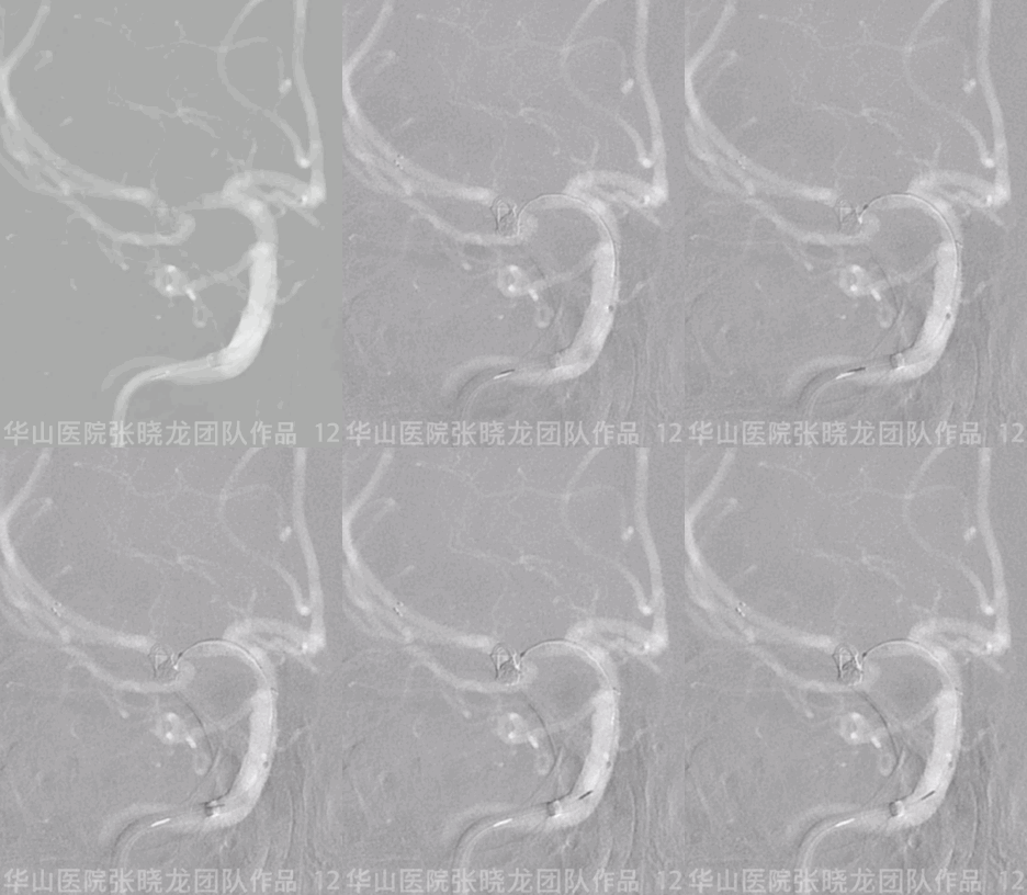
Figure 12 GIF. Released the tension of the coiling micro-catheter but failed to reinsert the loaded coil.
