Published in November 9, 2023
Large ophthalmic AN with LVIS-assisted large-coil embolization

Review

History

• 73 y/o, female.

• CC: Suffered from blurred vision for a month. Local hospital MRI revealed a left ophthalmic aneurysm ( not available) .

• NE: Identify finger count from 15cm, left temporal visual field defect ( without specialized eye exams). 

• Past medical history: Congenital deafness in the left ear; Cholecystectomy 7 years ago; Duodenal ulcer.
• Medication: (-)

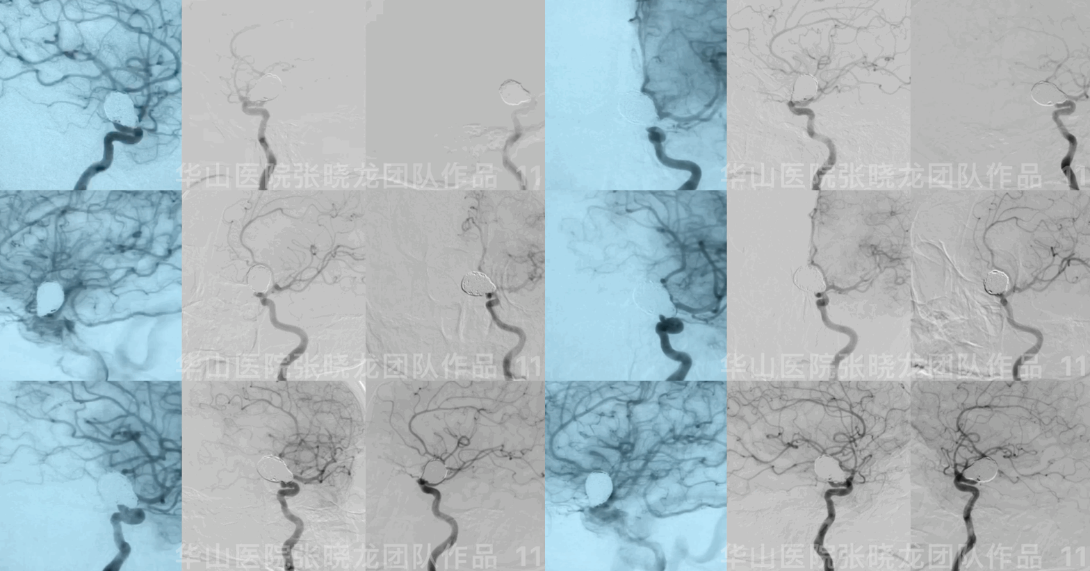
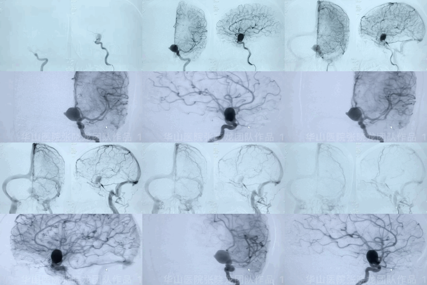
Figure 1 GIF. DSA reveals a large ophthalmic aneurysm with a daughter sac located in the upper-medial ICA wall. 


1

Treatment Strategy

• Irregular left large upper-medial ophthalmic aneurysm with mass effect should be treated. 

• Pipeline was unavailable at that time. So, the stent-assisted large coil technique was preferred to decrease the recurrence. 
• Block by Block embolization was the strategy for the case. And coil selection should be carefully considered– large coils for aneurysm sac to provide strong support and small coils with LVIS-stent assistance for aneurysm neck embolization to decrease neck relapse.

2

Operation

Figure 2 GIF.  Measurements. Large aneurysm with wide neck and daughter sac. Aneurysm sac: 16.9*15.5mm; daughter sac: 7.9mm; neck: 4.9mm. 


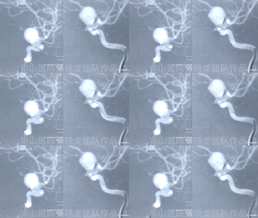
Figure 3 GIF. Microcatheter for coiling is navigated to the aneurysmal sac. 


Video 1. Microplex18 Cosmos Complex 20mm*65cm was inserted into the aneurysmal sac for framing. 


Figure 4 GIF. Insert the following coils to make the main sac densely packed: Three Microplex18 Cosmos Complex  16mm*52cm, Microplex18 Cosmos Complex 14mm*51cm. 


Video 2. Insert a Microplex18 Helical Regular 8mm*30cm coil.

视频 2. 填入1枚Microplex18 Helical Regular 8mm*30cm弹簧圈。
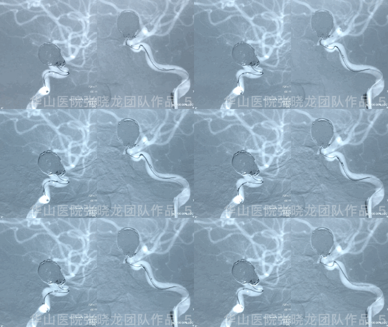

Video 3. Coils were inserted successively to pack the neck. During the coiling, repeated protrusion of the coil loop to the parent artery made it difficult to densely pack the neck.


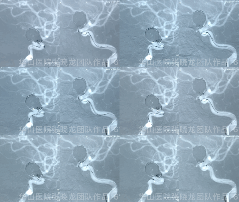
Figure 5 GIF. LVIS 3.5mm*20cm were deployed covering the aneurysmal sac. 


Figure 6 GIF. A Microplex10 Hypersoft Helical 3mm*8cm coil was inserted into the aneurysmal sac successfully without protrusion.


Figure 7 GIF. Then the coils inserted for dense packing of the neck were as followed: Microplex10 Hypersoft Helical 3mm*8cm, two; Microplex10 Hypersoft Helical 2mm*4cm, three; Microplex10 Hypersoft Helical 1.5mm*3cm, one; Microplex10 Hypersoft Helical 1mm*3cm, two.


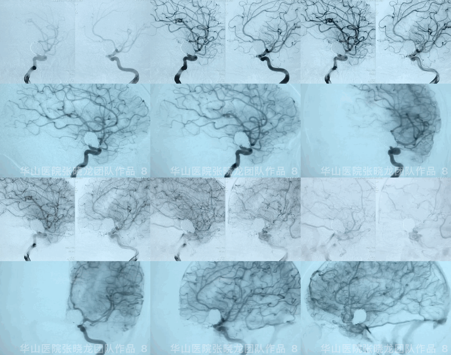
Figure 8 GIF. The aneurysm was densely packed with parent artery patent.


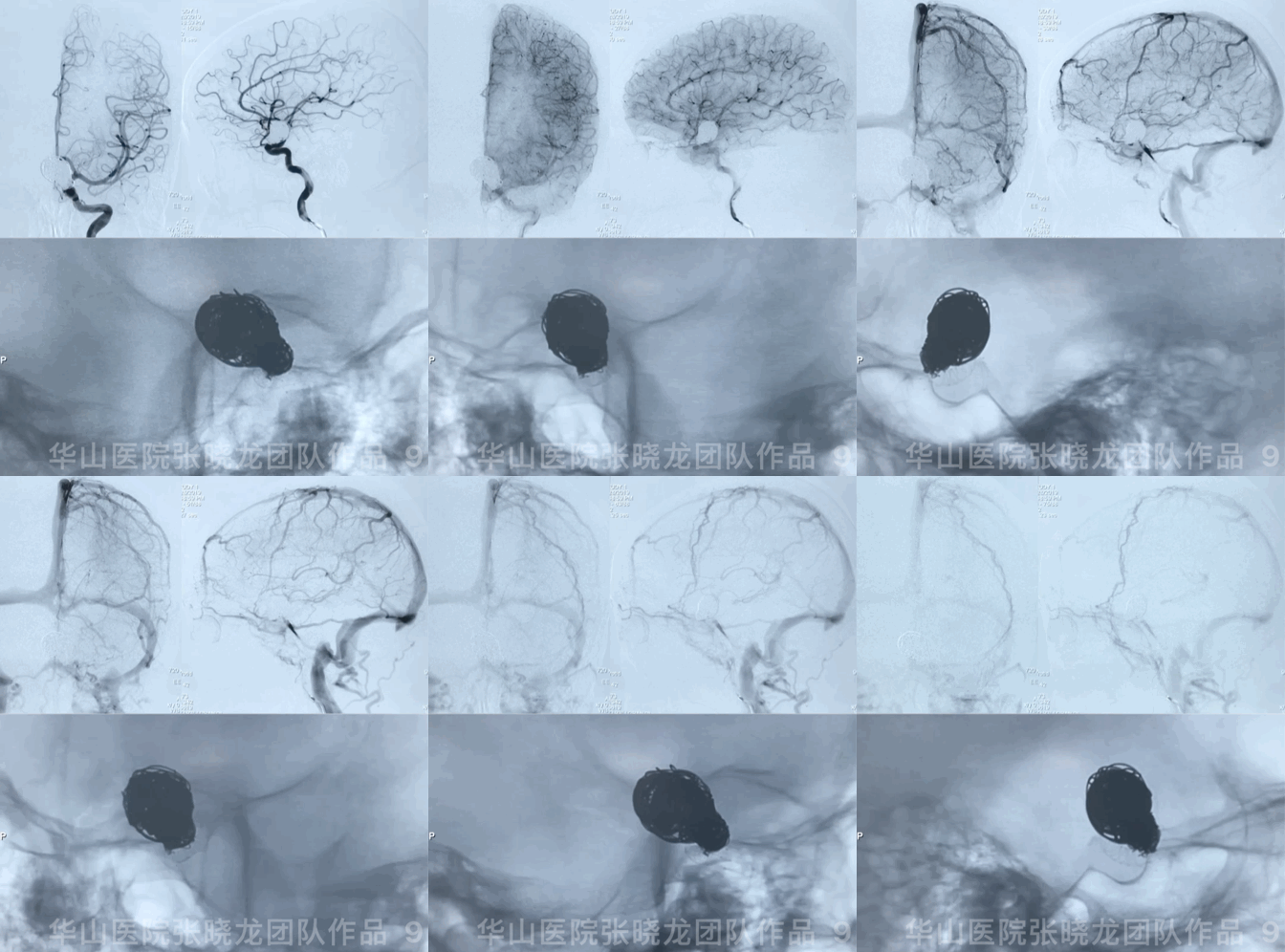
Figure 9 GIF. Intracranial vessels are intact. No evident hemorrhage or thrombus.

3

Post Operation

• GCS 15, left temporal visual field defect and blurred vision, no new neurological defect. 

• TEG: ADP 10.7%. 

• Medication: Suggest dual antiplatelets at least 3 months. However the patient stopped antiplatelets 2 months after operation.


4

Follow-up (4-month, 38-month)

• The patient still suffered from blurred vision and eye exams showed bilateral eye field defect. 

• Four-month and 38-month DSA revealed no residue or recurrence. 
• Next follow up is scheduled in 5 years.


Figure 10 GIF. Follow-up angiography shows no relapse of the aneurysm. 


Figure 11 GIF. Rotational DSA comparison among post-operation and twice follow up DSA shows complete occlusion of the aneurysm. 

6

Summary

  • Left large upper-medial ophthalmic aneurysm with mass effect should be treated. 

  • This case adopted the stent-assisted large coiling technique. Block by Block embolization was performed to pack the sac with large coils and embolize the neck with small coils, which can decrease the recurrence rate while the mass effect may remain existed.
  • Large coils for aneurysm sac provided strong support and LVIS assisted small coils for aneurysm neck embolization to decrease neck relapse. Large coiling technique could lower recurrence rate while mass effect may remain existed. 
  • A pipeline could be chosen to alleviate mass effect for this case. 
  • Coil selection is significant in this case. Though the first two coils did not provide a satisfied frame, the stable basket involving the neck was achieved by the third coil framing.
  • The right ophthalmic aneurysm is regular and small, indicating the low risk of rupture. It coud be follow-up.

Comments
Log into post your comments
1
2