Review
• 58 y/o, female.
• CC: Suffered from dizziness for 3 months. An anterior-wall ophthalmic aneurysm was detected from MRA in local hospital.
• Medical history: HTN for 10 years well-controlled. Meniere’s disease for 3 months.
• PE: (-)
Video 1. DSA shows a right irregular anterior-wall ophthalmic aneurysm with a daughter sac.
1
Strategy
• Since the irregular anterior-wall ophthalmic aneurysm has a daughter sac, the aneurysm has a high rupture risk due to flow impingement. Stent-assisted coil embolization is preferred for this wide-necked aneurysm.
• For the proximal tortuous route, Navien guiding catheter with a long-sheath is selected. General heparinization should be performed before navigating the guiding catheter.
• The daughter sac should be embolized to decrease the risk of delayed postoperative bleeding.
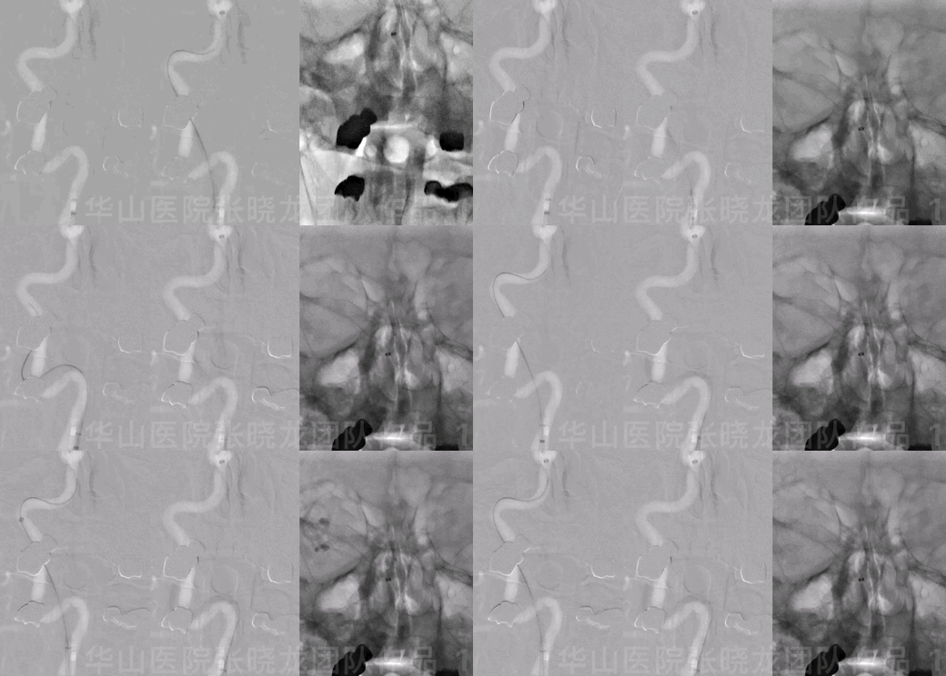
Figure 1 GIF. General heparinization 90cm long sheath+ 115cm 6F Navien.
1. The long-sheath was in the ICA proximal segment.
2. The guiding wire straightened the tortuous curve. Navien was in position.
3. Retrieve the guide wire and push the Navien guiding catheter complying to the previous ICA shape to prevent vasospasm and thrombosis.
4. Angiography confirmed no contrast medium stagnation.
2
Operation
Video 2. Angiography after guiding shows the thrombosis might have been formed.
Video 3. After placing a XT-27 microcatheter in the parent artery, an Echelon-10 45° shaped into an S curve was navigated to the aneurysmal sac. Neuroform 4*15mm was deployed via the XT-27 covering the aneurysmal neck.
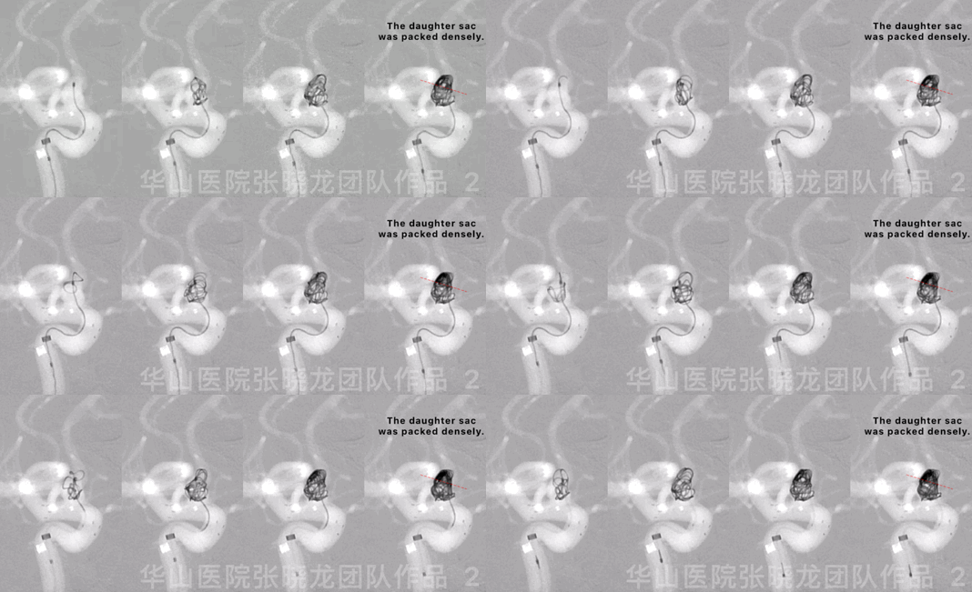
Figure 2 GIF. Four Target-360 4mm*8cm coils were inserted into the aneurysmal sac.
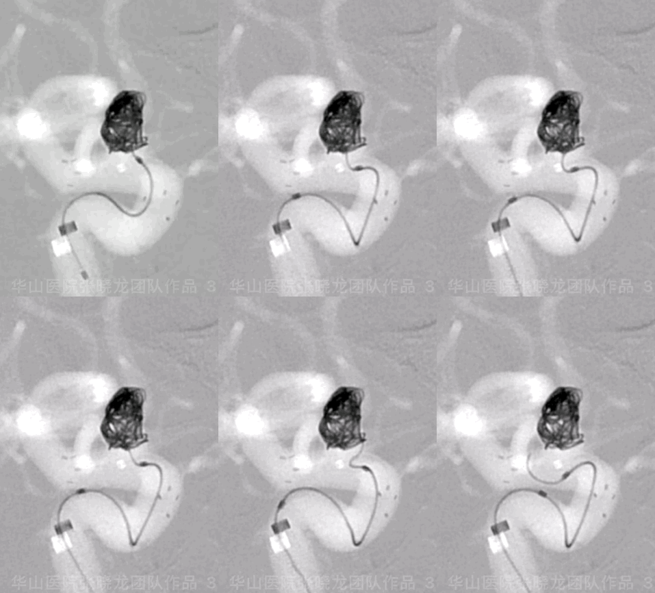
Figure 3 GIF. A Target-helical 4mm*8cm coil was unable to be inserted.
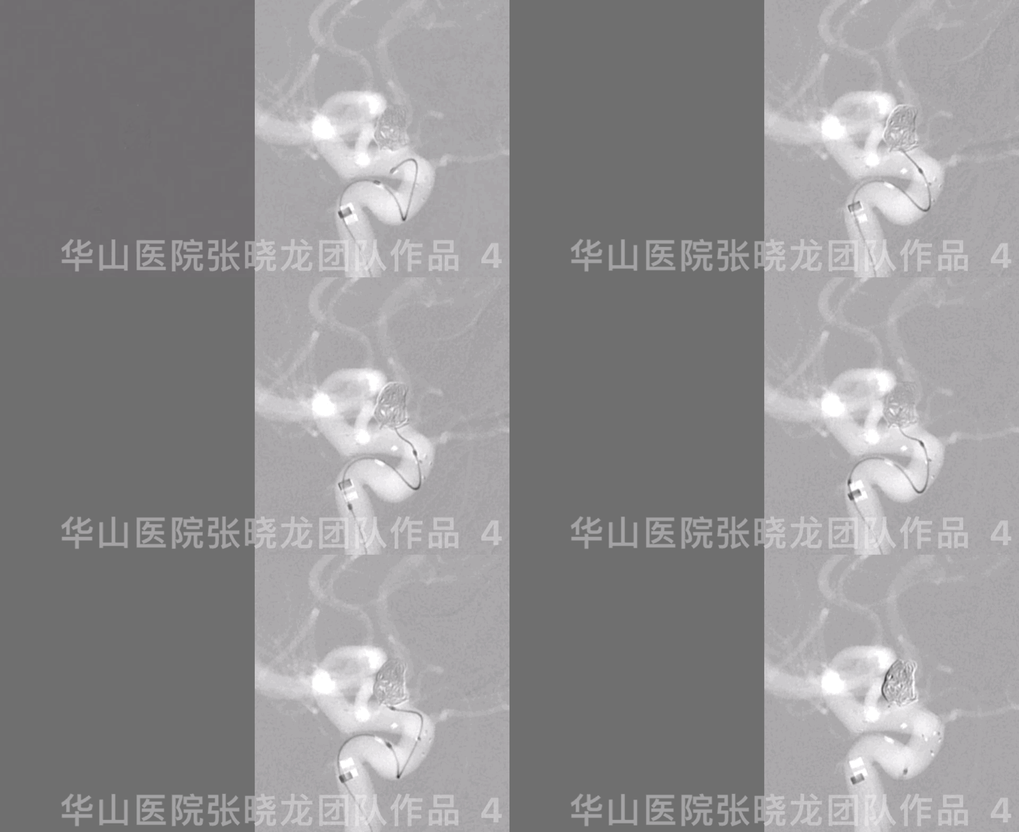
Figure 4 GIF. Renavigating the microcatheter into the aneurysm sac.
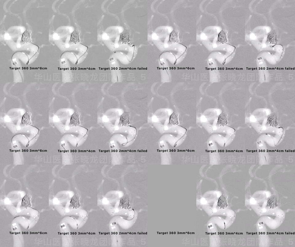
Figure 5 GIF. Another two Target-360 coils were inserted.
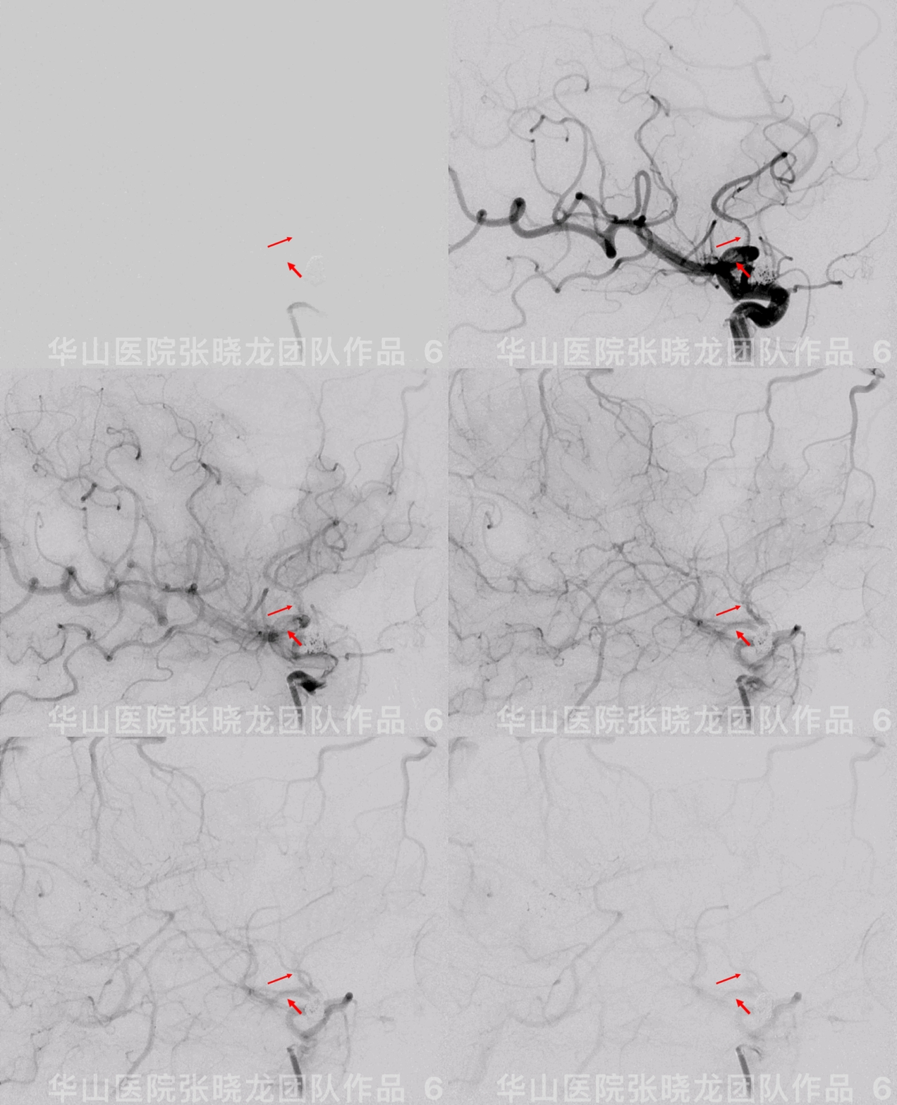
Figure 6 GIF. Thrombosis formed in the proximal MCA segment.
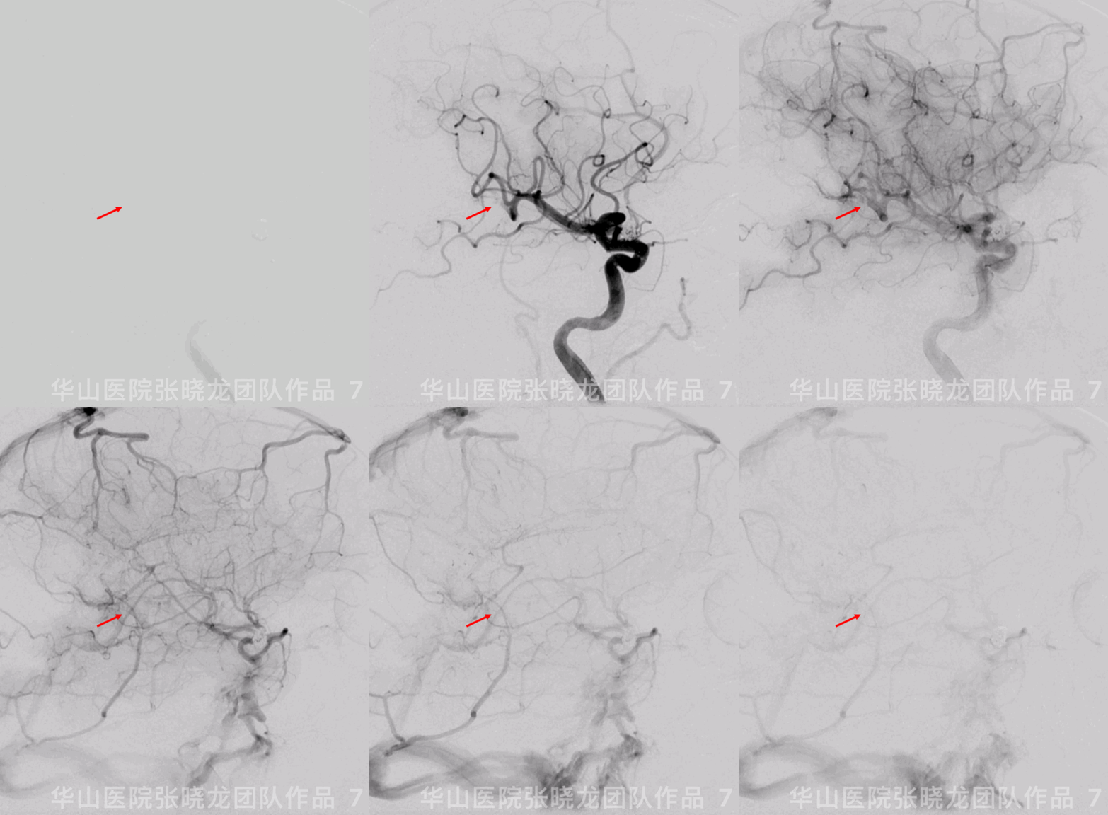
Figure 7 GIF. The thrombosis was flushed away into the parietal-occipital branch after retrieving the guiding catheter.

Figure 8 GIF. Improved blood flow of the right MCA after using Tirofiban 21ml (12ml+4ml+5ml) in 60 minutes. ACT: 207 sec, heparin 1ml.

Figure 9. Left VA angiography in the same working projection revealed that the ipsilateral PCA compensated the right parietal-occipital branch territory via the pia anastomosis.
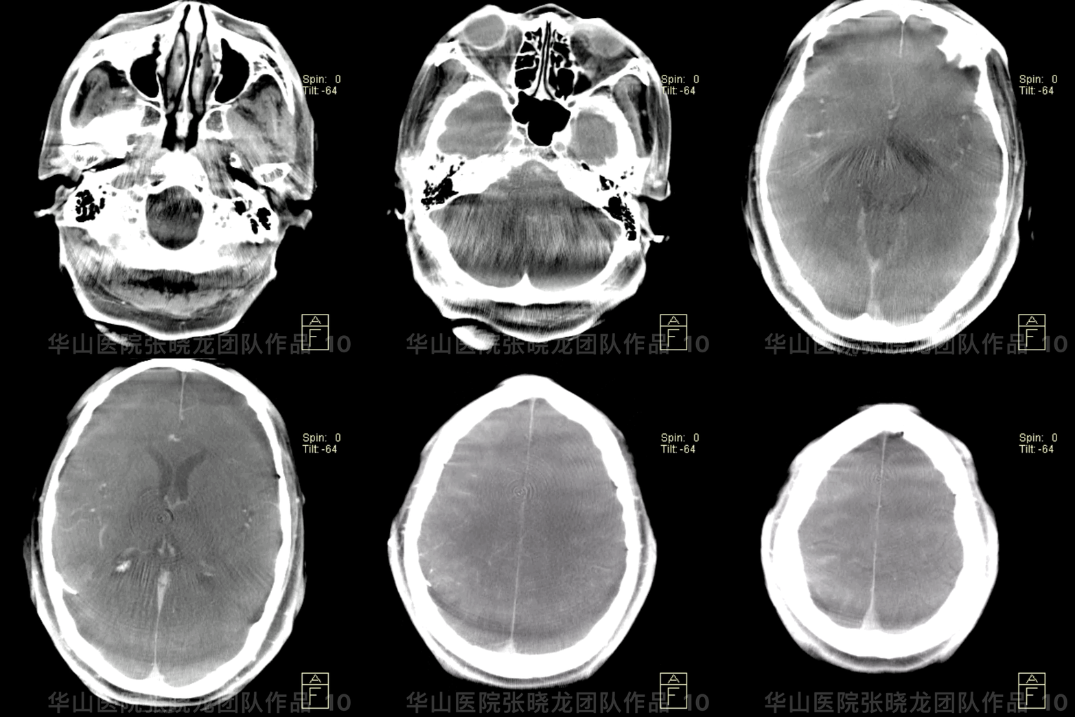
Figure 10 GIF. Post-operative DynaCT (-); PE (-)

Figure 11. CT 4 hours later: No hemorrhage or infarctions.

Figure 12. MRI DWI 48 hours later: Right temporal and occipital lobe infarction without significant symptoms, especially no visual deficiency.
3
Post Operation
• GCS 15, bilateral strength normal, bilateral pupil movement normal and light reflux normal, Babinskin negative.
• Tirofiban 8ml/h maintained 48 hours.
• ADP 55.7% and AA 93.1%
• At discharge: Clopidogrel for 3 months and Aspirin for long term.
4
Follow-up (9-month)

Figure 13 GIF. 9-month follow up angiography shows the precious occluded artery was patent.
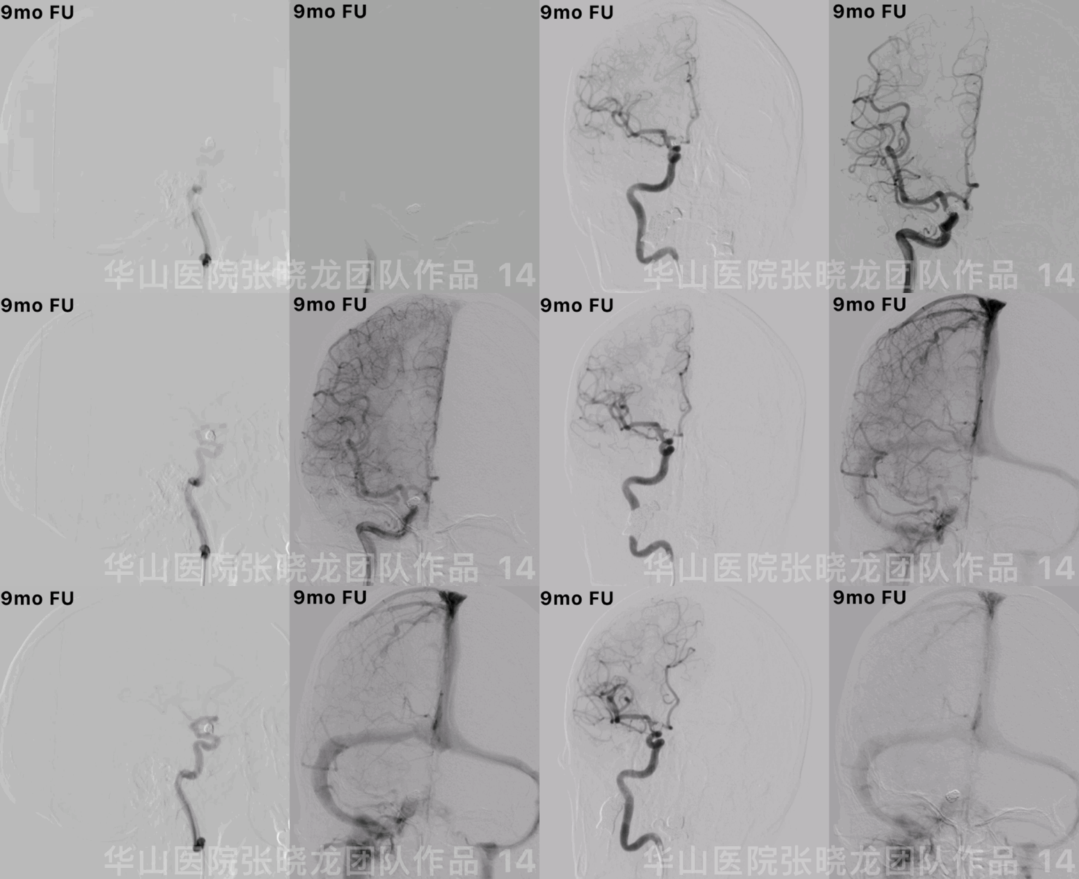
Figure 14 GIF. No relapse of the aneurysm. Stop Aspirin. Next follow up was scheduled in 2-3 years.
Summary
Since the irregular anterior-wall ophthalmic aneurysm has a daughter sac, the aneurysm has a high rupture risk due to flow impingement. Stent-assisted coil embolization is preferred for this wide-necked aneurysm.
For the proximal tortuous route, Navien guiding catheter with long-sheath is selected. General heparinization should be performed before navigating the guiding catheter.
1. The long-sheath was in the ICA proximal segment.
2. The guiding wire straightened the tortuous curve. Navien was in position.
3. Retrieve the guide wire and push the Navien guiding catheter conforming to the previous ICA shape.
4.Angiography confirmed no contrast medium stagnation.
The infusion system must be opened fluently in the long sheath in the ICA.
Large coiling technique was adopted to decrease recurrence risk because of the stable guiding catheter. The daughter sac should be embolized to decrease the delayed postoperative bleeding. When the thrombosis was flushed away into the parietal-occipital branch after retrieving the guiding catheter, Tirofiban was administered via the guiding catheter. Left VA angiography in the same working projection revealed that the ipsilateral PCA compensated the right parietal-occipital branch territory via the pia anastomosis.
